Magnesium »
PDB 2c42-2cic »
2c6y »
Magnesium in PDB 2c6y: Crystal Structure of Interleukin Enhancer-Binding Factor 1 Bound to Dna
Protein crystallography data
The structure of Crystal Structure of Interleukin Enhancer-Binding Factor 1 Bound to Dna, PDB code: 2c6y
was solved by
K.-L.Tsai,
C.-Y.Huang,
C.-H.Chang,
Y.-J.Sun,
W.-J.Chuang,
C.-D.Hsiao,
with X-Ray Crystallography technique. A brief refinement statistics is given in the table below:
| Resolution Low / High (Å) | 29.44 / 2.4 |
| Space group | P 61 2 2 |
| Cell size a, b, c (Å), α, β, γ (°) | 58.736, 58.736, 324.923, 90.00, 90.00, 120.00 |
| R / Rfree (%) | 23.3 / 25.8 |
Magnesium Binding Sites:
The binding sites of Magnesium atom in the Crystal Structure of Interleukin Enhancer-Binding Factor 1 Bound to Dna
(pdb code 2c6y). This binding sites where shown within
5.0 Angstroms radius around Magnesium atom.
In total 2 binding sites of Magnesium where determined in the Crystal Structure of Interleukin Enhancer-Binding Factor 1 Bound to Dna, PDB code: 2c6y:
Jump to Magnesium binding site number: 1; 2;
In total 2 binding sites of Magnesium where determined in the Crystal Structure of Interleukin Enhancer-Binding Factor 1 Bound to Dna, PDB code: 2c6y:
Jump to Magnesium binding site number: 1; 2;
Magnesium binding site 1 out of 2 in 2c6y
Go back to
Magnesium binding site 1 out
of 2 in the Crystal Structure of Interleukin Enhancer-Binding Factor 1 Bound to Dna
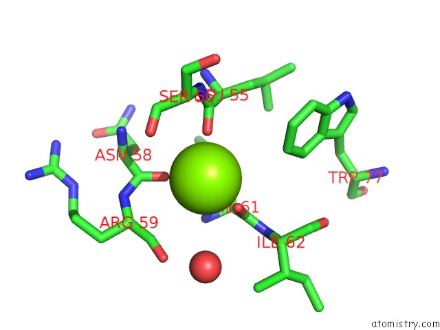
Mono view
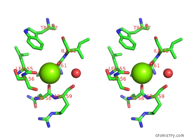
Stereo pair view
Mono view
Stereo pair view
A full contact list of Magnesium with other atoms in the Mg binding
site number 1 of Crystal Structure of Interleukin Enhancer-Binding Factor 1 Bound to Dna within 5.0Å range:
|
Magnesium binding site 2 out of 2 in 2c6y
Go back to
Magnesium binding site 2 out
of 2 in the Crystal Structure of Interleukin Enhancer-Binding Factor 1 Bound to Dna
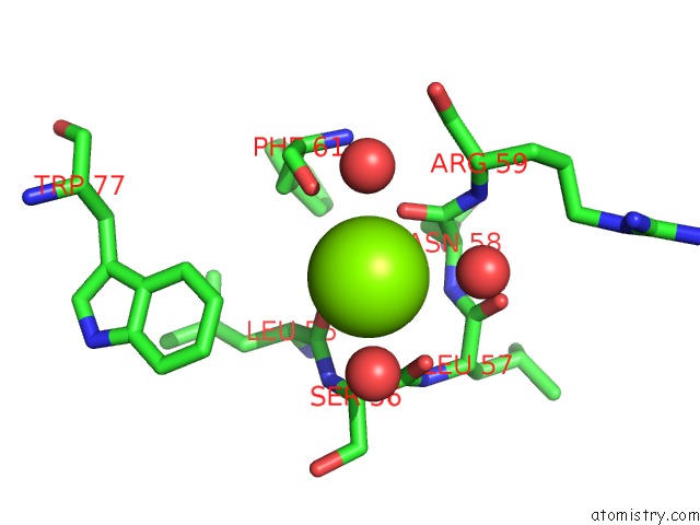
Mono view
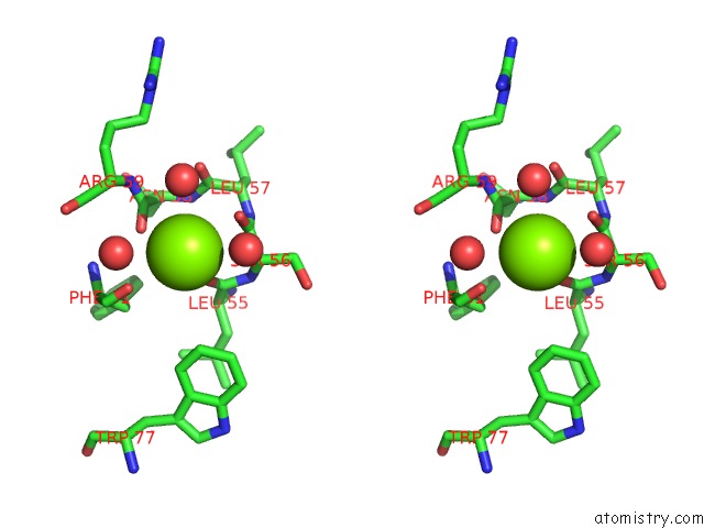
Stereo pair view
Mono view
Stereo pair view
A full contact list of Magnesium with other atoms in the Mg binding
site number 2 of Crystal Structure of Interleukin Enhancer-Binding Factor 1 Bound to Dna within 5.0Å range:
|
Reference:
K.-L.Tsai,
C.-Y.Huang,
C.-H.Chang,
Y.-J.Sun,
W.-J.Chuang,
C.-D.Hsiao.
Crystal Structure of the Human FOXK1A-Dna Complex and Its Implications on the Diverse Binding Specificity of Winged Helix/Forkhead Proteins. J.Biol.Chem. V. 281 17400 2006.
ISSN: ISSN 0021-9258
PubMed: 16624804
DOI: 10.1074/JBC.M600478200
Page generated: Tue Aug 13 22:12:47 2024
ISSN: ISSN 0021-9258
PubMed: 16624804
DOI: 10.1074/JBC.M600478200
Last articles
F in 7GRXF in 7H3Z
F in 7H3Y
F in 7H3O
F in 7H34
F in 7H2L
F in 7H1K
F in 7GS5
F in 7GSC
F in 7GRN