Magnesium »
PDB 2c42-2cic »
2cfr »
Magnesium in PDB 2cfr: Crystal Structure of Human Pyridoxal 5'-Phosphate Phosphatase
Enzymatic activity of Crystal Structure of Human Pyridoxal 5'-Phosphate Phosphatase
All present enzymatic activity of Crystal Structure of Human Pyridoxal 5'-Phosphate Phosphatase:
3.1.3.74;
3.1.3.74;
Protein crystallography data
The structure of Crystal Structure of Human Pyridoxal 5'-Phosphate Phosphatase, PDB code: 2cfr
was solved by
B.S.Kang,
H.J.Cho,
K.J.Kim,
O.S.Kwon,
with X-Ray Crystallography technique. A brief refinement statistics is given in the table below:
| Resolution Low / High (Å) | 19.06 / 2.4 |
| Space group | P 43 21 2 |
| Cell size a, b, c (Å), α, β, γ (°) | 54.300, 54.300, 213.900, 90.00, 90.00, 90.00 |
| R / Rfree (%) | 20.3 / 25.7 |
Magnesium Binding Sites:
The binding sites of Magnesium atom in the Crystal Structure of Human Pyridoxal 5'-Phosphate Phosphatase
(pdb code 2cfr). This binding sites where shown within
5.0 Angstroms radius around Magnesium atom.
In total only one binding site of Magnesium was determined in the Crystal Structure of Human Pyridoxal 5'-Phosphate Phosphatase, PDB code: 2cfr:
In total only one binding site of Magnesium was determined in the Crystal Structure of Human Pyridoxal 5'-Phosphate Phosphatase, PDB code: 2cfr:
Magnesium binding site 1 out of 1 in 2cfr
Go back to
Magnesium binding site 1 out
of 1 in the Crystal Structure of Human Pyridoxal 5'-Phosphate Phosphatase
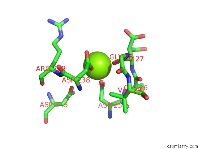
Mono view
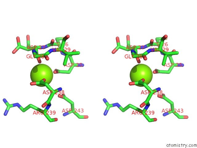
Stereo pair view
Mono view
Stereo pair view
A full contact list of Magnesium with other atoms in the Mg binding
site number 1 of Crystal Structure of Human Pyridoxal 5'-Phosphate Phosphatase within 5.0Å range:
|
Reference:
B.S.Kang,
H.J.Cho,
K.J.Kim,
O.S.Kwon.
Crystal Structure of Human Pyridoxal 5'-Phosphate Phosphatase To Be Published.
Page generated: Sun Aug 10 10:16:30 2025
Last articles
Mg in 3CCUMg in 3CCS
Mg in 3CCR
Mg in 3CCQ
Mg in 3CCM
Mg in 3CCL
Mg in 3CCJ
Mg in 3CC2
Mg in 3CCE
Mg in 3CC7