Magnesium »
PDB 2c42-2cic »
2che »
Magnesium in PDB 2che: Structure of the MG2+-Bound Form of Chey and Mechanism of Phosphoryl Transfer in Bacterial Chemotaxis
Protein crystallography data
The structure of Structure of the MG2+-Bound Form of Chey and Mechanism of Phosphoryl Transfer in Bacterial Chemotaxis, PDB code: 2che
was solved by
A.Stock,
E.Martinez-Hackert,
B.Rasmussen,
A.West,
J.Stock,
D.Ringe,
G.Petsko,
with X-Ray Crystallography technique. A brief refinement statistics is given in the table below:
| Resolution Low / High (Å) | N/A / 1.80 |
| Space group | P 21 21 21 |
| Cell size a, b, c (Å), α, β, γ (°) | 45.710, 46.980, 53.910, 90.00, 90.00, 90.00 |
| R / Rfree (%) | n/a / n/a |
Magnesium Binding Sites:
The binding sites of Magnesium atom in the Structure of the MG2+-Bound Form of Chey and Mechanism of Phosphoryl Transfer in Bacterial Chemotaxis
(pdb code 2che). This binding sites where shown within
5.0 Angstroms radius around Magnesium atom.
In total only one binding site of Magnesium was determined in the Structure of the MG2+-Bound Form of Chey and Mechanism of Phosphoryl Transfer in Bacterial Chemotaxis, PDB code: 2che:
In total only one binding site of Magnesium was determined in the Structure of the MG2+-Bound Form of Chey and Mechanism of Phosphoryl Transfer in Bacterial Chemotaxis, PDB code: 2che:
Magnesium binding site 1 out of 1 in 2che
Go back to
Magnesium binding site 1 out
of 1 in the Structure of the MG2+-Bound Form of Chey and Mechanism of Phosphoryl Transfer in Bacterial Chemotaxis
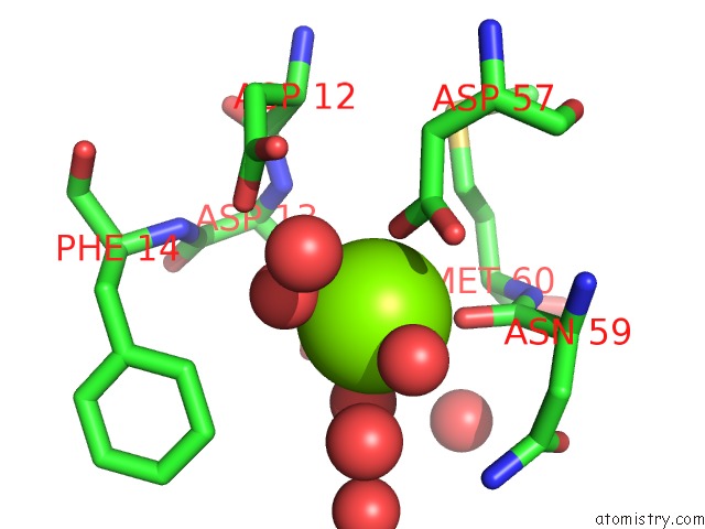
Mono view
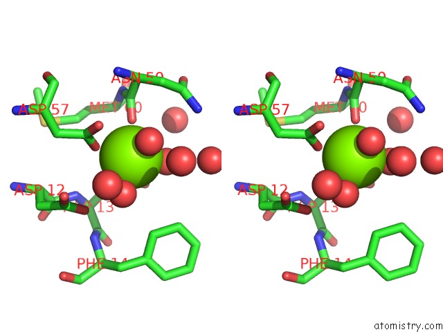
Stereo pair view
Mono view
Stereo pair view
A full contact list of Magnesium with other atoms in the Mg binding
site number 1 of Structure of the MG2+-Bound Form of Chey and Mechanism of Phosphoryl Transfer in Bacterial Chemotaxis within 5.0Å range:
|
Reference:
A.M.Stock,
E.Martinez-Hackert,
B.F.Rasmussen,
A.H.West,
J.B.Stock,
D.Ringe,
G.A.Petsko.
Structure of the Mg(2+)-Bound Form of Chey and Mechanism of Phosphoryl Transfer in Bacterial Chemotaxis. Biochemistry V. 32 13375 1993.
ISSN: ISSN 0006-2960
PubMed: 8257674
DOI: 10.1021/BI00212A001
Page generated: Sun Aug 10 10:17:09 2025
ISSN: ISSN 0006-2960
PubMed: 8257674
DOI: 10.1021/BI00212A001
Last articles
Mg in 3CMTMg in 3CIS
Mg in 3CMR
Mg in 3CMQ
Mg in 3CLY
Mg in 3CK5
Mg in 3CLC
Mg in 3CKG
Mg in 3CKE
Mg in 3CK4