Magnesium »
PDB 2cie-2cvy »
2cje »
Magnesium in PDB 2cje: The Crystal Structure of A Complex of Leishmania Major Dutpase with Substrate Analogue Dupnhp
Enzymatic activity of The Crystal Structure of A Complex of Leishmania Major Dutpase with Substrate Analogue Dupnhp
All present enzymatic activity of The Crystal Structure of A Complex of Leishmania Major Dutpase with Substrate Analogue Dupnhp:
3.6.1.23;
3.6.1.23;
Protein crystallography data
The structure of The Crystal Structure of A Complex of Leishmania Major Dutpase with Substrate Analogue Dupnhp, PDB code: 2cje
was solved by
O.V.Moroz,
M.J.Fogg,
D.Gonzalez-Pacanowska,
K.S.Wilson,
with X-Ray Crystallography technique. A brief refinement statistics is given in the table below:
| Resolution Low / High (Å) | 76.03 / 2.34 |
| Space group | P 65 2 2 |
| Cell size a, b, c (Å), α, β, γ (°) | 87.866, 87.866, 146.531, 90.00, 90.00, 120.00 |
| R / Rfree (%) | 18.4 / 23.8 |
Magnesium Binding Sites:
The binding sites of Magnesium atom in the The Crystal Structure of A Complex of Leishmania Major Dutpase with Substrate Analogue Dupnhp
(pdb code 2cje). This binding sites where shown within
5.0 Angstroms radius around Magnesium atom.
In total 3 binding sites of Magnesium where determined in the The Crystal Structure of A Complex of Leishmania Major Dutpase with Substrate Analogue Dupnhp, PDB code: 2cje:
Jump to Magnesium binding site number: 1; 2; 3;
In total 3 binding sites of Magnesium where determined in the The Crystal Structure of A Complex of Leishmania Major Dutpase with Substrate Analogue Dupnhp, PDB code: 2cje:
Jump to Magnesium binding site number: 1; 2; 3;
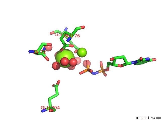
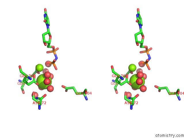
Magnesium binding site 1 out of 3 in 2cje
Go back to
Magnesium binding site 1 out
of 3 in the The Crystal Structure of A Complex of Leishmania Major Dutpase with Substrate Analogue Dupnhp
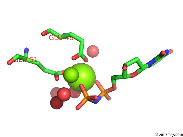
Mono view
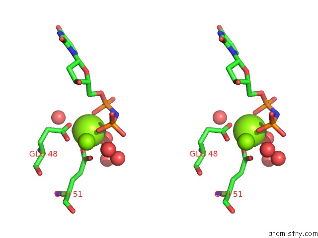
Stereo pair view
Mono view
Stereo pair view
A full contact list of Magnesium with other atoms in the Mg binding
site number 1 of The Crystal Structure of A Complex of Leishmania Major Dutpase with Substrate Analogue Dupnhp within 5.0Å range:
|
Magnesium binding site 2 out of 3 in 2cje
Go back to
Magnesium binding site 2 out
of 3 in the The Crystal Structure of A Complex of Leishmania Major Dutpase with Substrate Analogue Dupnhp
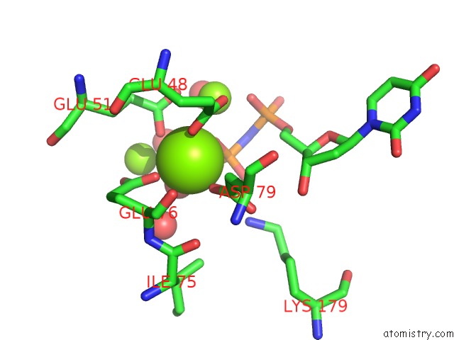
Mono view
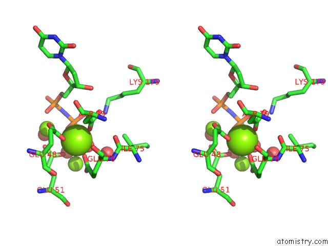
Stereo pair view
Mono view
Stereo pair view
A full contact list of Magnesium with other atoms in the Mg binding
site number 2 of The Crystal Structure of A Complex of Leishmania Major Dutpase with Substrate Analogue Dupnhp within 5.0Å range:
|
Magnesium binding site 3 out of 3 in 2cje
Go back to
Magnesium binding site 3 out
of 3 in the The Crystal Structure of A Complex of Leishmania Major Dutpase with Substrate Analogue Dupnhp
Mono view
Stereo pair view
Mono view
Stereo pair view
A full contact list of Magnesium with other atoms in the Mg binding
site number 3 of The Crystal Structure of A Complex of Leishmania Major Dutpase with Substrate Analogue Dupnhp within 5.0Å range:
|
Reference:
G.R.Hemsworth,
O.V.Moroz,
M.J.Fogg,
B.Scott,
C.Bosch-Navarrete,
D.Gonzalez-Pacanowska,
K.S.Wilson.
The Crystal Structure of the Leishmania Major Deoxyuridine Triphosphate Nucleotidohydrolase in Complex with Nucleotide Analogues, Dump, and Deoxyuridine. J.Biol.Chem. V. 286 16470 2011.
ISSN: ISSN 0021-9258
PubMed: 21454646
DOI: 10.1074/JBC.M111.224873
Page generated: Tue Aug 13 22:17:10 2024
ISSN: ISSN 0021-9258
PubMed: 21454646
DOI: 10.1074/JBC.M111.224873
Last articles
Zn in 9J0NZn in 9J0O
Zn in 9J0P
Zn in 9FJX
Zn in 9EKB
Zn in 9C0F
Zn in 9CAH
Zn in 9CH0
Zn in 9CH3
Zn in 9CH1