Magnesium »
PDB 2cie-2cvy »
2cl7 »
Magnesium in PDB 2cl7: Crystal Structure Analysis of A Fluorescent Form of H-Ras P21 in Complex with Gtp
Protein crystallography data
The structure of Crystal Structure Analysis of A Fluorescent Form of H-Ras P21 in Complex with Gtp, PDB code: 2cl7
was solved by
B.U.Klink,
R.S.Goody,
A.J.Scheidig,
with X-Ray Crystallography technique. A brief refinement statistics is given in the table below:
| Resolution Low / High (Å) | 69.34 / 1.25 |
| Space group | P 41 |
| Cell size a, b, c (Å), α, β, γ (°) | 69.280, 69.280, 35.020, 90.00, 90.00, 90.00 |
| R / Rfree (%) | 14.8 / 17 |
Magnesium Binding Sites:
The binding sites of Magnesium atom in the Crystal Structure Analysis of A Fluorescent Form of H-Ras P21 in Complex with Gtp
(pdb code 2cl7). This binding sites where shown within
5.0 Angstroms radius around Magnesium atom.
In total 2 binding sites of Magnesium where determined in the Crystal Structure Analysis of A Fluorescent Form of H-Ras P21 in Complex with Gtp, PDB code: 2cl7:
Jump to Magnesium binding site number: 1; 2;
In total 2 binding sites of Magnesium where determined in the Crystal Structure Analysis of A Fluorescent Form of H-Ras P21 in Complex with Gtp, PDB code: 2cl7:
Jump to Magnesium binding site number: 1; 2;
Magnesium binding site 1 out of 2 in 2cl7
Go back to
Magnesium binding site 1 out
of 2 in the Crystal Structure Analysis of A Fluorescent Form of H-Ras P21 in Complex with Gtp
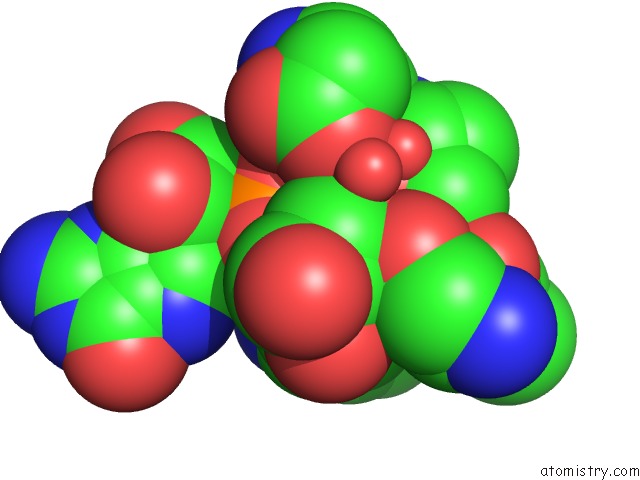
Mono view
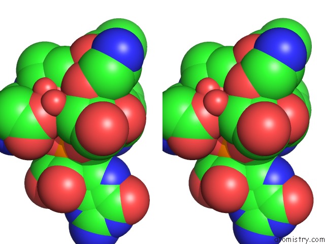
Stereo pair view
Mono view
Stereo pair view
A full contact list of Magnesium with other atoms in the Mg binding
site number 1 of Crystal Structure Analysis of A Fluorescent Form of H-Ras P21 in Complex with Gtp within 5.0Å range:
|
Magnesium binding site 2 out of 2 in 2cl7
Go back to
Magnesium binding site 2 out
of 2 in the Crystal Structure Analysis of A Fluorescent Form of H-Ras P21 in Complex with Gtp
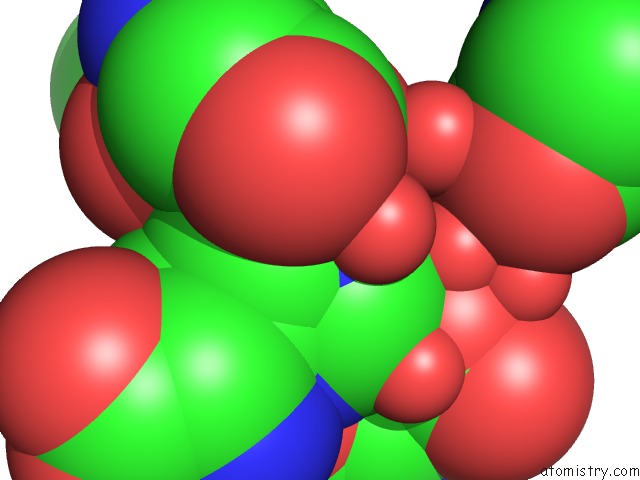
Mono view
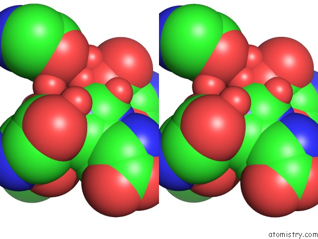
Stereo pair view
Mono view
Stereo pair view
A full contact list of Magnesium with other atoms in the Mg binding
site number 2 of Crystal Structure Analysis of A Fluorescent Form of H-Ras P21 in Complex with Gtp within 5.0Å range:
|
Reference:
B.U.Klink,
R.S.Goody,
A.J.Scheidig.
A Newly Designed Microspectrofluorometer For Kinetic Studies on Protein Crystals in Combination with X-Ray Diffraction. Biophys.J. V. 91 981 2006.
ISSN: ISSN 0006-3495
PubMed: 16698776
DOI: 10.1529/BIOPHYSJ.105.078931
Page generated: Tue Aug 13 22:18:19 2024
ISSN: ISSN 0006-3495
PubMed: 16698776
DOI: 10.1529/BIOPHYSJ.105.078931
Last articles
Zn in 9MJ5Zn in 9HNW
Zn in 9G0L
Zn in 9FNE
Zn in 9DZN
Zn in 9E0I
Zn in 9D32
Zn in 9DAK
Zn in 8ZXC
Zn in 8ZUF