Magnesium »
PDB 2dlc-2e74 »
2dpc »
Magnesium in PDB 2dpc: Crystal Structure of D(Cgcgaatxcgcg) Where X Is 5-(N- Aminohexyl)Carbamoyl-2'-O-Methyluridine
Protein crystallography data
The structure of Crystal Structure of D(Cgcgaatxcgcg) Where X Is 5-(N- Aminohexyl)Carbamoyl-2'-O-Methyluridine, PDB code: 2dpc
was solved by
E.C.M.Juan,
J.Kondo,
T.Kurihara,
T.Ito,
Y.Ueno,
A.Matsuda,
A.Takenaka,
with X-Ray Crystallography technique. A brief refinement statistics is given in the table below:
| Resolution Low / High (Å) | 10.00 / 1.55 |
| Space group | P 21 21 21 |
| Cell size a, b, c (Å), α, β, γ (°) | 25.214, 40.533, 63.887, 90.00, 90.00, 90.00 |
| R / Rfree (%) | 21.5 / 26.6 |
Other elements in 2dpc:
The structure of Crystal Structure of D(Cgcgaatxcgcg) Where X Is 5-(N- Aminohexyl)Carbamoyl-2'-O-Methyluridine also contains other interesting chemical elements:
| Cobalt | (Co) | 3 atoms |
Magnesium Binding Sites:
The binding sites of Magnesium atom in the Crystal Structure of D(Cgcgaatxcgcg) Where X Is 5-(N- Aminohexyl)Carbamoyl-2'-O-Methyluridine
(pdb code 2dpc). This binding sites where shown within
5.0 Angstroms radius around Magnesium atom.
In total only one binding site of Magnesium was determined in the Crystal Structure of D(Cgcgaatxcgcg) Where X Is 5-(N- Aminohexyl)Carbamoyl-2'-O-Methyluridine, PDB code: 2dpc:
In total only one binding site of Magnesium was determined in the Crystal Structure of D(Cgcgaatxcgcg) Where X Is 5-(N- Aminohexyl)Carbamoyl-2'-O-Methyluridine, PDB code: 2dpc:
Magnesium binding site 1 out of 1 in 2dpc
Go back to
Magnesium binding site 1 out
of 1 in the Crystal Structure of D(Cgcgaatxcgcg) Where X Is 5-(N- Aminohexyl)Carbamoyl-2'-O-Methyluridine
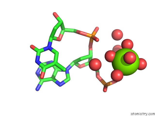
Mono view
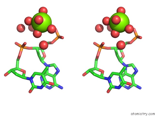
Stereo pair view
Mono view
Stereo pair view
A full contact list of Magnesium with other atoms in the Mg binding
site number 1 of Crystal Structure of D(Cgcgaatxcgcg) Where X Is 5-(N- Aminohexyl)Carbamoyl-2'-O-Methyluridine within 5.0Å range:
|
Reference:
E.C.M.Juan,
J.Kondo,
T.Kurihara,
T.Ito,
Y.Ueno,
A.Matsuda,
A.Takenaka.
Crystal Structures of Dna:Dna and Dna:Rna Duplexes Containing 5-(N-Aminohexyl)Carbamoyl-Modified Uracils Reveal the Basis For Properties As Antigene and Antisense Molecules Nucleic Acids Res. V. 35 1969 2007.
ISSN: ISSN 0305-1048
PubMed: 17341465
DOI: 10.1093/NAR/GKL821
Page generated: Tue Aug 13 22:34:04 2024
ISSN: ISSN 0305-1048
PubMed: 17341465
DOI: 10.1093/NAR/GKL821
Last articles
Zn in 9MJ5Zn in 9HNW
Zn in 9G0L
Zn in 9FNE
Zn in 9DZN
Zn in 9E0I
Zn in 9D32
Zn in 9DAK
Zn in 8ZXC
Zn in 8ZUF