Magnesium »
PDB 2dlc-2e74 »
2e2q »
Magnesium in PDB 2e2q: Crystal Structure of Sulfolobus Tokodaii Hexokinase in Complex with Xylose, MG2+, and Adp
Enzymatic activity of Crystal Structure of Sulfolobus Tokodaii Hexokinase in Complex with Xylose, MG2+, and Adp
All present enzymatic activity of Crystal Structure of Sulfolobus Tokodaii Hexokinase in Complex with Xylose, MG2+, and Adp:
2.7.1.1;
2.7.1.1;
Protein crystallography data
The structure of Crystal Structure of Sulfolobus Tokodaii Hexokinase in Complex with Xylose, MG2+, and Adp, PDB code: 2e2q
was solved by
H.Nishimasu,
S.Fushinobu,
H.Shoun,
T.Wakagi,
with X-Ray Crystallography technique. A brief refinement statistics is given in the table below:
| Resolution Low / High (Å) | 49.53 / 2.00 |
| Space group | C 1 2 1 |
| Cell size a, b, c (Å), α, β, γ (°) | 143.440, 81.470, 52.510, 90.00, 109.40, 90.00 |
| R / Rfree (%) | 21.3 / 25.5 |
Magnesium Binding Sites:
The binding sites of Magnesium atom in the Crystal Structure of Sulfolobus Tokodaii Hexokinase in Complex with Xylose, MG2+, and Adp
(pdb code 2e2q). This binding sites where shown within
5.0 Angstroms radius around Magnesium atom.
In total only one binding site of Magnesium was determined in the Crystal Structure of Sulfolobus Tokodaii Hexokinase in Complex with Xylose, MG2+, and Adp, PDB code: 2e2q:
In total only one binding site of Magnesium was determined in the Crystal Structure of Sulfolobus Tokodaii Hexokinase in Complex with Xylose, MG2+, and Adp, PDB code: 2e2q:
Magnesium binding site 1 out of 1 in 2e2q
Go back to
Magnesium binding site 1 out
of 1 in the Crystal Structure of Sulfolobus Tokodaii Hexokinase in Complex with Xylose, MG2+, and Adp
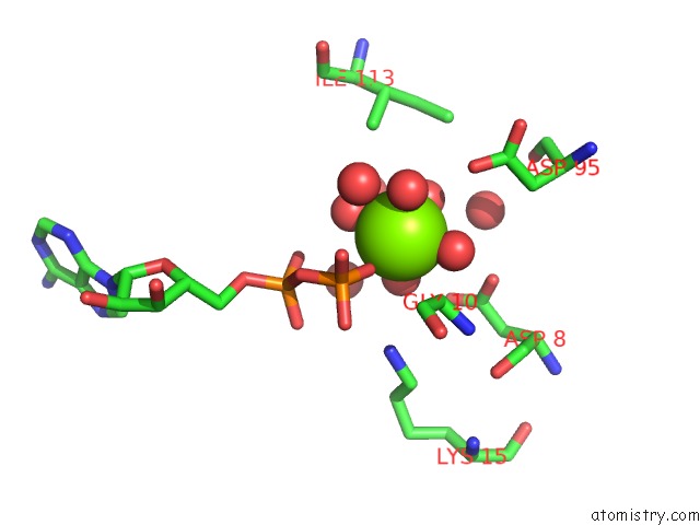
Mono view
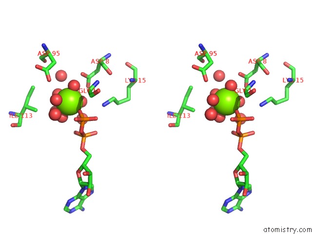
Stereo pair view
Mono view
Stereo pair view
A full contact list of Magnesium with other atoms in the Mg binding
site number 1 of Crystal Structure of Sulfolobus Tokodaii Hexokinase in Complex with Xylose, MG2+, and Adp within 5.0Å range:
|
Reference:
H.Nishimasu,
S.Fushinobu,
H.Shoun,
T.Wakagi.
Crystal Structures of An Atp-Dependent Hexokinase with Broad Substrate Specificity From the Hyperthermophilic Archaeon Sulfolobus Tokodaii. J.Biol.Chem. V. 282 9923 2007.
ISSN: ISSN 0021-9258
PubMed: 17229727
DOI: 10.1074/JBC.M610678200
Page generated: Tue Aug 13 22:39:10 2024
ISSN: ISSN 0021-9258
PubMed: 17229727
DOI: 10.1074/JBC.M610678200
Last articles
Fe in 2YXOFe in 2YRS
Fe in 2YXC
Fe in 2YNM
Fe in 2YVJ
Fe in 2YP1
Fe in 2YU2
Fe in 2YU1
Fe in 2YQB
Fe in 2YOO