Magnesium »
PDB 2e75-2ein »
2eb6 »
Magnesium in PDB 2eb6: Crystal Structure of Hpcg Complexed with Mg Ion
Protein crystallography data
The structure of Crystal Structure of Hpcg Complexed with Mg Ion, PDB code: 2eb6
was solved by
A.Izumi,
D.Rea,
T.Adachi,
S.Unzai,
S.Y.Park,
D.I.Roper,
J.R.H.Tame,
with X-Ray Crystallography technique. A brief refinement statistics is given in the table below:
| Resolution Low / High (Å) | 10.00 / 1.69 |
| Space group | P 43 21 2 |
| Cell size a, b, c (Å), α, β, γ (°) | 135.955, 135.955, 194.064, 90.00, 90.00, 90.00 |
| R / Rfree (%) | 19.4 / 21.3 |
Magnesium Binding Sites:
The binding sites of Magnesium atom in the Crystal Structure of Hpcg Complexed with Mg Ion
(pdb code 2eb6). This binding sites where shown within
5.0 Angstroms radius around Magnesium atom.
In total 5 binding sites of Magnesium where determined in the Crystal Structure of Hpcg Complexed with Mg Ion, PDB code: 2eb6:
Jump to Magnesium binding site number: 1; 2; 3; 4; 5;
In total 5 binding sites of Magnesium where determined in the Crystal Structure of Hpcg Complexed with Mg Ion, PDB code: 2eb6:
Jump to Magnesium binding site number: 1; 2; 3; 4; 5;
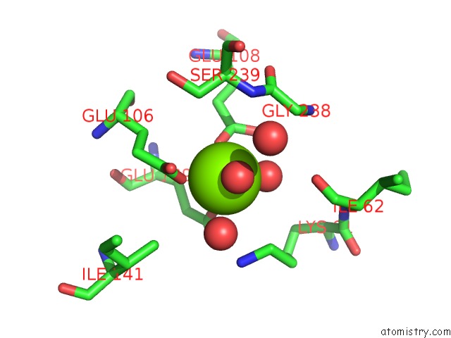
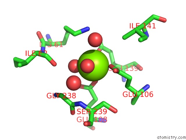
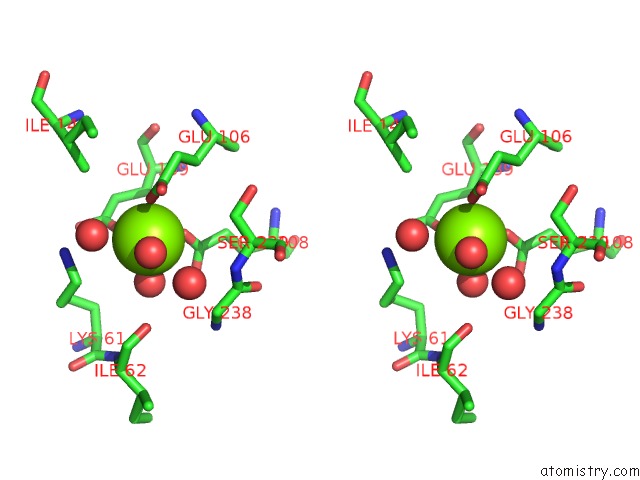
Magnesium binding site 1 out of 5 in 2eb6
Go back to
Magnesium binding site 1 out
of 5 in the Crystal Structure of Hpcg Complexed with Mg Ion
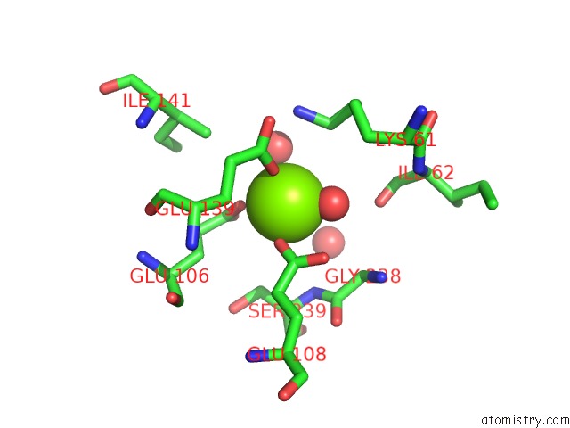
Mono view
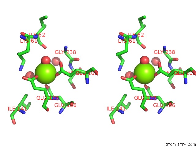
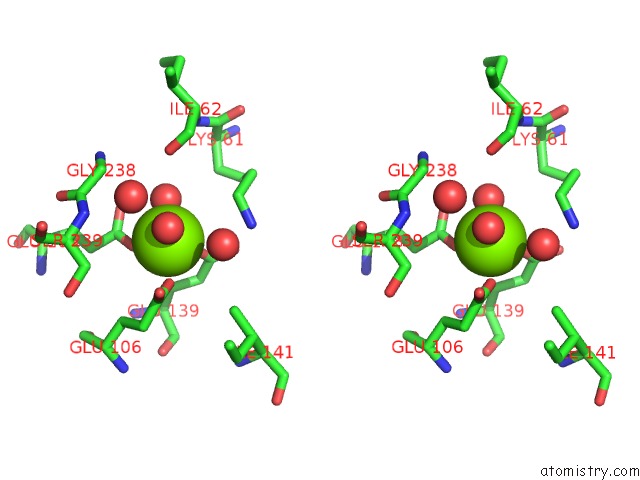
Stereo pair view
Mono view
Stereo pair view
A full contact list of Magnesium with other atoms in the Mg binding
site number 1 of Crystal Structure of Hpcg Complexed with Mg Ion within 5.0Å range:
|
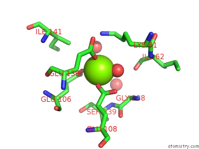
Magnesium binding site 2 out of 5 in 2eb6
Go back to
Magnesium binding site 2 out
of 5 in the Crystal Structure of Hpcg Complexed with Mg Ion
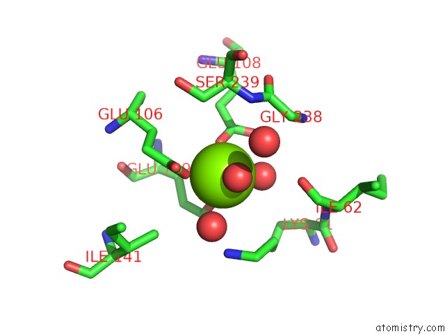
Mono view
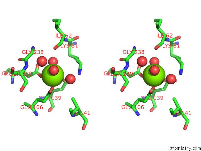
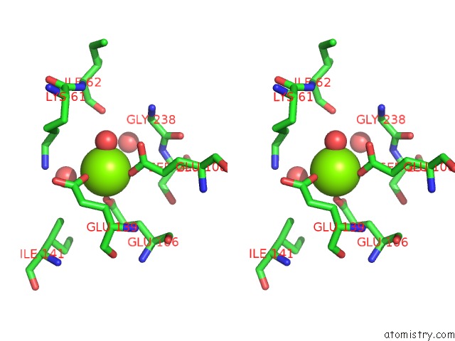
Stereo pair view
Mono view
Stereo pair view
A full contact list of Magnesium with other atoms in the Mg binding
site number 2 of Crystal Structure of Hpcg Complexed with Mg Ion within 5.0Å range:
|
Magnesium binding site 3 out of 5 in 2eb6
Go back to
Magnesium binding site 3 out
of 5 in the Crystal Structure of Hpcg Complexed with Mg Ion
Mono view
Stereo pair view
Mono view
Stereo pair view
A full contact list of Magnesium with other atoms in the Mg binding
site number 3 of Crystal Structure of Hpcg Complexed with Mg Ion within 5.0Å range:
|
Magnesium binding site 4 out of 5 in 2eb6
Go back to
Magnesium binding site 4 out
of 5 in the Crystal Structure of Hpcg Complexed with Mg Ion
Mono view
Stereo pair view
Mono view
Stereo pair view
A full contact list of Magnesium with other atoms in the Mg binding
site number 4 of Crystal Structure of Hpcg Complexed with Mg Ion within 5.0Å range:
|
Magnesium binding site 5 out of 5 in 2eb6
Go back to
Magnesium binding site 5 out
of 5 in the Crystal Structure of Hpcg Complexed with Mg Ion
Mono view
Stereo pair view
Mono view
Stereo pair view
A full contact list of Magnesium with other atoms in the Mg binding
site number 5 of Crystal Structure of Hpcg Complexed with Mg Ion within 5.0Å range:
|
Reference:
A.Izumi,
D.Rea,
T.Adachi,
S.Unzai,
S.Y.Park,
D.I.Roper,
J.R.H.Tame.
Structure and Mechanism of Hpcg, A Hydratase in the Homoprotocatechuate Degradation Pathway of Escherichia Coli J.Mol.Biol. V. 370 899 2007.
ISSN: ISSN 0022-2836
PubMed: 17559873
DOI: 10.1016/J.JMB.2007.05.006
Page generated: Tue Aug 13 22:44:31 2024
ISSN: ISSN 0022-2836
PubMed: 17559873
DOI: 10.1016/J.JMB.2007.05.006
Last articles
Cl in 5WZWCl in 5X0Q
Cl in 5X0V
Cl in 5WZV
Cl in 5X08
Cl in 5WZO
Cl in 5WZU
Cl in 5WZM
Cl in 5WWV
Cl in 5WZS