Magnesium »
PDB 2e75-2ein »
2ei1 »
Magnesium in PDB 2ei1: Anaerobic Crystal Strucutre Analysis of the 1,2-Dihydroxynaphthalene Dioxygeanse of Pseudomonas Sp. Strain C18 Complexes to 1,2- Dihydroxynaphthalene
Protein crystallography data
The structure of Anaerobic Crystal Strucutre Analysis of the 1,2-Dihydroxynaphthalene Dioxygeanse of Pseudomonas Sp. Strain C18 Complexes to 1,2- Dihydroxynaphthalene, PDB code: 2ei1
was solved by
D.B.Neau,
M.S.Kelker,
C.L.Colbert,
J.T.Bolin,
with X-Ray Crystallography technique. A brief refinement statistics is given in the table below:
| Resolution Low / High (Å) | 50.00 / 1.52 |
| Space group | I 4 2 2 |
| Cell size a, b, c (Å), α, β, γ (°) | 117.848, 117.848, 120.965, 90.00, 90.00, 90.00 |
| R / Rfree (%) | 17.1 / 18 |
Other elements in 2ei1:
The structure of Anaerobic Crystal Strucutre Analysis of the 1,2-Dihydroxynaphthalene Dioxygeanse of Pseudomonas Sp. Strain C18 Complexes to 1,2- Dihydroxynaphthalene also contains other interesting chemical elements:
| Iron | (Fe) | 1 atom |
Magnesium Binding Sites:
The binding sites of Magnesium atom in the Anaerobic Crystal Strucutre Analysis of the 1,2-Dihydroxynaphthalene Dioxygeanse of Pseudomonas Sp. Strain C18 Complexes to 1,2- Dihydroxynaphthalene
(pdb code 2ei1). This binding sites where shown within
5.0 Angstroms radius around Magnesium atom.
In total only one binding site of Magnesium was determined in the Anaerobic Crystal Strucutre Analysis of the 1,2-Dihydroxynaphthalene Dioxygeanse of Pseudomonas Sp. Strain C18 Complexes to 1,2- Dihydroxynaphthalene, PDB code: 2ei1:
In total only one binding site of Magnesium was determined in the Anaerobic Crystal Strucutre Analysis of the 1,2-Dihydroxynaphthalene Dioxygeanse of Pseudomonas Sp. Strain C18 Complexes to 1,2- Dihydroxynaphthalene, PDB code: 2ei1:
Magnesium binding site 1 out of 1 in 2ei1
Go back to
Magnesium binding site 1 out
of 1 in the Anaerobic Crystal Strucutre Analysis of the 1,2-Dihydroxynaphthalene Dioxygeanse of Pseudomonas Sp. Strain C18 Complexes to 1,2- Dihydroxynaphthalene
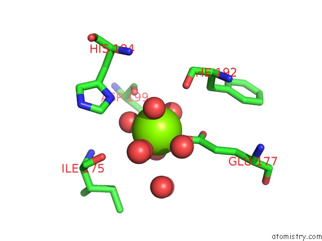
Mono view
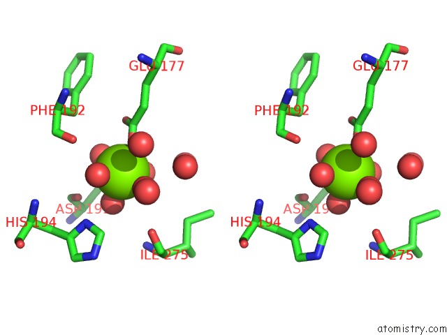
Stereo pair view
Mono view
Stereo pair view
A full contact list of Magnesium with other atoms in the Mg binding
site number 1 of Anaerobic Crystal Strucutre Analysis of the 1,2-Dihydroxynaphthalene Dioxygeanse of Pseudomonas Sp. Strain C18 Complexes to 1,2- Dihydroxynaphthalene within 5.0Å range:
|
Reference:
D.B.Neau,
M.S.Kelker,
H.Maaroufi,
C.L.Colbert,
L.D.Eltis,
J.T.Bolin.
Structural Explanation For Success and Failure in the Enzymatic Ring-Cleavage of 3,4 Dihydroxybiphenyl and Related Pcb Metabolites. To Be Published.
Page generated: Tue Aug 13 22:46:36 2024
Last articles
Zn in 9JYWZn in 9IR4
Zn in 9IR3
Zn in 9GMX
Zn in 9GMW
Zn in 9JEJ
Zn in 9ERF
Zn in 9ERE
Zn in 9EGV
Zn in 9EGW