Magnesium »
PDB 2e75-2ein »
2eim »
Magnesium in PDB 2eim: Zinc Ion Binding Structure of Bovine Heart Cytochrome C Oxidase in the Fully Reduced State
Enzymatic activity of Zinc Ion Binding Structure of Bovine Heart Cytochrome C Oxidase in the Fully Reduced State
All present enzymatic activity of Zinc Ion Binding Structure of Bovine Heart Cytochrome C Oxidase in the Fully Reduced State:
1.9.3.1;
1.9.3.1;
Protein crystallography data
The structure of Zinc Ion Binding Structure of Bovine Heart Cytochrome C Oxidase in the Fully Reduced State, PDB code: 2eim
was solved by
K.Muramoto,
K.Hirata,
K.Shinzawa-Itoh,
S.Yoko-O,
E.Yamashita,
H.Aoyama,
T.Tsukihara,
S.Yoshikawa,
with X-Ray Crystallography technique. A brief refinement statistics is given in the table below:
| Resolution Low / High (Å) | 40.00 / 2.60 |
| Space group | P 21 21 21 |
| Cell size a, b, c (Å), α, β, γ (°) | 183.909, 206.721, 178.337, 90.00, 90.00, 90.00 |
| R / Rfree (%) | 20.4 / 25.6 |
Other elements in 2eim:
The structure of Zinc Ion Binding Structure of Bovine Heart Cytochrome C Oxidase in the Fully Reduced State also contains other interesting chemical elements:
| Zinc | (Zn) | 6 atoms |
| Iron | (Fe) | 4 atoms |
| Copper | (Cu) | 6 atoms |
| Sodium | (Na) | 2 atoms |
Magnesium Binding Sites:
The binding sites of Magnesium atom in the Zinc Ion Binding Structure of Bovine Heart Cytochrome C Oxidase in the Fully Reduced State
(pdb code 2eim). This binding sites where shown within
5.0 Angstroms radius around Magnesium atom.
In total 2 binding sites of Magnesium where determined in the Zinc Ion Binding Structure of Bovine Heart Cytochrome C Oxidase in the Fully Reduced State, PDB code: 2eim:
Jump to Magnesium binding site number: 1; 2;
In total 2 binding sites of Magnesium where determined in the Zinc Ion Binding Structure of Bovine Heart Cytochrome C Oxidase in the Fully Reduced State, PDB code: 2eim:
Jump to Magnesium binding site number: 1; 2;
Magnesium binding site 1 out of 2 in 2eim
Go back to
Magnesium binding site 1 out
of 2 in the Zinc Ion Binding Structure of Bovine Heart Cytochrome C Oxidase in the Fully Reduced State
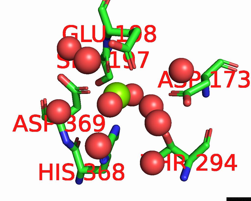
Mono view
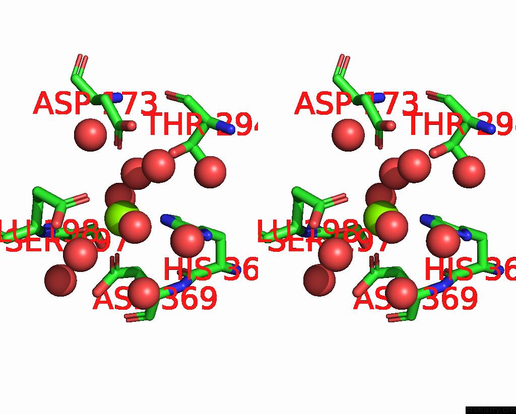
Stereo pair view
Mono view
Stereo pair view
A full contact list of Magnesium with other atoms in the Mg binding
site number 1 of Zinc Ion Binding Structure of Bovine Heart Cytochrome C Oxidase in the Fully Reduced State within 5.0Å range:
|
Magnesium binding site 2 out of 2 in 2eim
Go back to
Magnesium binding site 2 out
of 2 in the Zinc Ion Binding Structure of Bovine Heart Cytochrome C Oxidase in the Fully Reduced State
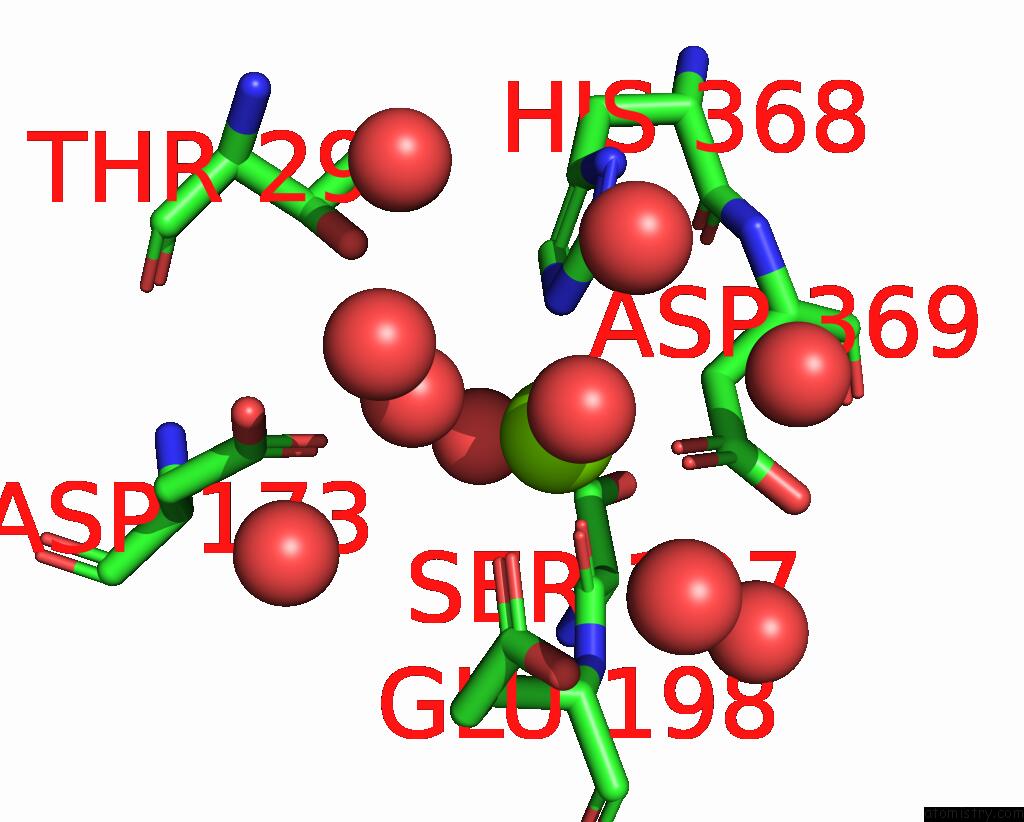
Mono view
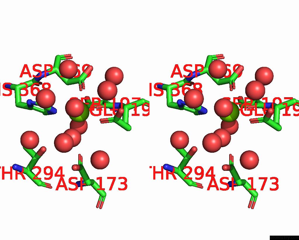
Stereo pair view
Mono view
Stereo pair view
A full contact list of Magnesium with other atoms in the Mg binding
site number 2 of Zinc Ion Binding Structure of Bovine Heart Cytochrome C Oxidase in the Fully Reduced State within 5.0Å range:
|
Reference:
K.Muramoto,
K.Hirata,
K.Shinzawa-Itoh,
S.Yoko-O,
E.Yamashita,
H.Aoyama,
T.Tsukihara,
S.Yoshikawa.
A Histidine Residue Acting As A Controlling Site For Dioxygen Reduction and Proton Pumping By Cytochrome C Oxidase Proc.Natl.Acad.Sci.Usa V. 104 7881 2007.
ISSN: ISSN 0027-8424
PubMed: 17470809
DOI: 10.1073/PNAS.0610031104
Page generated: Tue Aug 13 22:47:03 2024
ISSN: ISSN 0027-8424
PubMed: 17470809
DOI: 10.1073/PNAS.0610031104
Last articles
Ca in 5ONQCa in 5OLS
Ca in 5OLP
Ca in 5OLQ
Ca in 5OL2
Ca in 5OLB
Ca in 5OL7
Ca in 5OFX
Ca in 5ODU
Ca in 5OHT