Magnesium »
PDB 2f6y-2fl2 »
2f9l »
Magnesium in PDB 2f9l: 3D Structure of Inactive Human RAB11B Gtpase
Enzymatic activity of 3D Structure of Inactive Human RAB11B Gtpase
All present enzymatic activity of 3D Structure of Inactive Human RAB11B Gtpase:
3.6.5.2;
3.6.5.2;
Protein crystallography data
The structure of 3D Structure of Inactive Human RAB11B Gtpase, PDB code: 2f9l
was solved by
S.M.N.Scapin,
B.G.Guimaraes,
N.I.T.Zanchin,
with X-Ray Crystallography technique. A brief refinement statistics is given in the table below:
| Resolution Low / High (Å) | 39.22 / 1.55 |
| Space group | P 21 21 21 |
| Cell size a, b, c (Å), α, β, γ (°) | 45.790, 52.218, 59.351, 90.00, 90.00, 90.00 |
| R / Rfree (%) | 18.6 / 23.8 |
Magnesium Binding Sites:
The binding sites of Magnesium atom in the 3D Structure of Inactive Human RAB11B Gtpase
(pdb code 2f9l). This binding sites where shown within
5.0 Angstroms radius around Magnesium atom.
In total only one binding site of Magnesium was determined in the 3D Structure of Inactive Human RAB11B Gtpase, PDB code: 2f9l:
In total only one binding site of Magnesium was determined in the 3D Structure of Inactive Human RAB11B Gtpase, PDB code: 2f9l:
Magnesium binding site 1 out of 1 in 2f9l
Go back to
Magnesium binding site 1 out
of 1 in the 3D Structure of Inactive Human RAB11B Gtpase
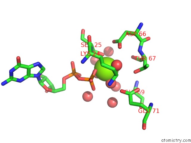
Mono view
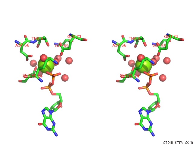
Stereo pair view
Mono view
Stereo pair view
A full contact list of Magnesium with other atoms in the Mg binding
site number 1 of 3D Structure of Inactive Human RAB11B Gtpase within 5.0Å range:
|
Reference:
S.M.Scapin,
F.R.Carneiro,
A.C.Alves,
F.J.Medrano,
B.G.Guimaraes,
N.I.Zanchin.
The Crystal Structure of the Small Gtpase RAB11B Reveals Critical Differences Relative to the RAB11A Isoform. J.Struct.Biol. V. 154 260 2006.
ISSN: ISSN 1047-8477
PubMed: 16545962
DOI: 10.1016/J.JSB.2006.01.007
Page generated: Tue Aug 13 23:05:18 2024
ISSN: ISSN 1047-8477
PubMed: 16545962
DOI: 10.1016/J.JSB.2006.01.007
Last articles
Zn in 9J0NZn in 9J0O
Zn in 9J0P
Zn in 9FJX
Zn in 9EKB
Zn in 9C0F
Zn in 9CAH
Zn in 9CH0
Zn in 9CH3
Zn in 9CH1