Magnesium »
PDB 2f6y-2fl2 »
2f9r »
Magnesium in PDB 2f9r: Crystal Structure of the Inactive State of the Smase I, A Sphingomyelinase D From Loxosceles Laeta Venom
Enzymatic activity of Crystal Structure of the Inactive State of the Smase I, A Sphingomyelinase D From Loxosceles Laeta Venom
All present enzymatic activity of Crystal Structure of the Inactive State of the Smase I, A Sphingomyelinase D From Loxosceles Laeta Venom:
3.1.4.41;
3.1.4.41;
Protein crystallography data
The structure of Crystal Structure of the Inactive State of the Smase I, A Sphingomyelinase D From Loxosceles Laeta Venom, PDB code: 2f9r
was solved by
M.T.Murakami,
A.Gabdoulkhakov,
M.F.Fernandes-Pedrosa,
C.Betzel,
D.V.Tambourgi,
R.K.Arni,
with X-Ray Crystallography technique. A brief refinement statistics is given in the table below:
| Resolution Low / High (Å) | 20.00 / 1.85 |
| Space group | P 65 |
| Cell size a, b, c (Å), α, β, γ (°) | 140.587, 140.587, 113.608, 90.00, 90.00, 120.00 |
| R / Rfree (%) | 18.7 / 23.4 |
Magnesium Binding Sites:
The binding sites of Magnesium atom in the Crystal Structure of the Inactive State of the Smase I, A Sphingomyelinase D From Loxosceles Laeta Venom
(pdb code 2f9r). This binding sites where shown within
5.0 Angstroms radius around Magnesium atom.
In total 4 binding sites of Magnesium where determined in the Crystal Structure of the Inactive State of the Smase I, A Sphingomyelinase D From Loxosceles Laeta Venom, PDB code: 2f9r:
Jump to Magnesium binding site number: 1; 2; 3; 4;
In total 4 binding sites of Magnesium where determined in the Crystal Structure of the Inactive State of the Smase I, A Sphingomyelinase D From Loxosceles Laeta Venom, PDB code: 2f9r:
Jump to Magnesium binding site number: 1; 2; 3; 4;
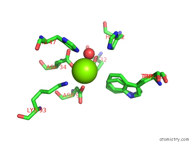
Magnesium binding site 1 out of 4 in 2f9r
Go back to
Magnesium binding site 1 out
of 4 in the Crystal Structure of the Inactive State of the Smase I, A Sphingomyelinase D From Loxosceles Laeta Venom

Mono view
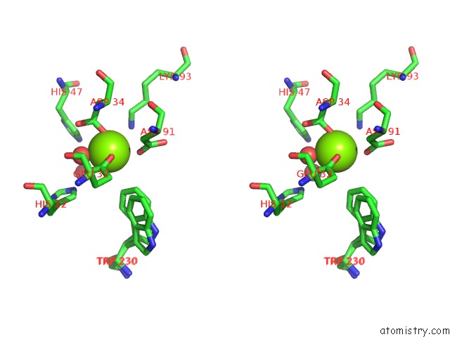
Stereo pair view
Mono view
Stereo pair view
A full contact list of Magnesium with other atoms in the Mg binding
site number 1 of Crystal Structure of the Inactive State of the Smase I, A Sphingomyelinase D From Loxosceles Laeta Venom within 5.0Å range:
|
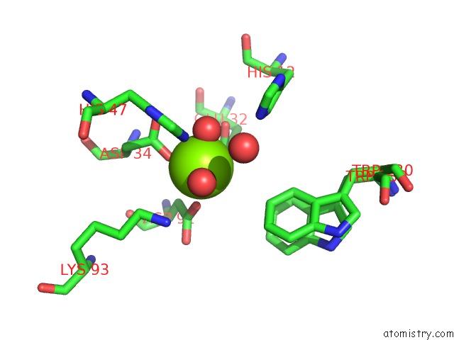
Magnesium binding site 2 out of 4 in 2f9r
Go back to
Magnesium binding site 2 out
of 4 in the Crystal Structure of the Inactive State of the Smase I, A Sphingomyelinase D From Loxosceles Laeta Venom
Mono view

Stereo pair view
Mono view
Stereo pair view
A full contact list of Magnesium with other atoms in the Mg binding
site number 2 of Crystal Structure of the Inactive State of the Smase I, A Sphingomyelinase D From Loxosceles Laeta Venom within 5.0Å range:
|
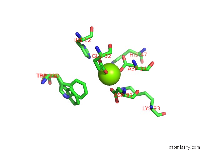
Magnesium binding site 3 out of 4 in 2f9r
Go back to
Magnesium binding site 3 out
of 4 in the Crystal Structure of the Inactive State of the Smase I, A Sphingomyelinase D From Loxosceles Laeta Venom
Mono view
Stereo pair view
Mono view
Stereo pair view
A full contact list of Magnesium with other atoms in the Mg binding
site number 3 of Crystal Structure of the Inactive State of the Smase I, A Sphingomyelinase D From Loxosceles Laeta Venom within 5.0Å range:
|
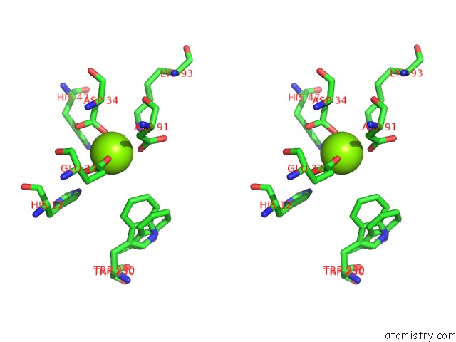
Magnesium binding site 4 out of 4 in 2f9r
Go back to
Magnesium binding site 4 out
of 4 in the Crystal Structure of the Inactive State of the Smase I, A Sphingomyelinase D From Loxosceles Laeta Venom
Mono view

Stereo pair view
Mono view
Stereo pair view
A full contact list of Magnesium with other atoms in the Mg binding
site number 4 of Crystal Structure of the Inactive State of the Smase I, A Sphingomyelinase D From Loxosceles Laeta Venom within 5.0Å range:
|
Reference:
M.T.Murakami,
A.Gabdoulkhakov,
M.F.Fernandes-Pedrosa,
D.V.Tambourgi,
R.K.Arni.
Structural Basis For Metal Ion Coordination and the Catalytic Mechanism of Sphingomyelinases D. J.Biol.Chem. V. 280 13658 2005.
ISSN: ISSN 0021-9258
PubMed: 15654080
DOI: 10.1074/JBC.M412437200
Page generated: Tue Aug 13 23:05:48 2024
ISSN: ISSN 0021-9258
PubMed: 15654080
DOI: 10.1074/JBC.M412437200
Last articles
F in 4ENHF in 4EPX
F in 4ENC
F in 4ENB
F in 4EMV
F in 4ENA
F in 4EN5
F in 4EKC
F in 4EKD
F in 4EHG