Magnesium »
PDB 2f6y-2fl2 »
2fgc »
Magnesium in PDB 2fgc: Crystal Structure of Acetolactate Synthase- Small Subunit From Thermotoga Maritima
Enzymatic activity of Crystal Structure of Acetolactate Synthase- Small Subunit From Thermotoga Maritima
All present enzymatic activity of Crystal Structure of Acetolactate Synthase- Small Subunit From Thermotoga Maritima:
2.2.1.6;
2.2.1.6;
Protein crystallography data
The structure of Crystal Structure of Acetolactate Synthase- Small Subunit From Thermotoga Maritima, PDB code: 2fgc
was solved by
J.J.Petkowski,
M.Chruszcz,
M.D.Zimmerman,
H.Zheng,
M.T.Cymborowski,
K.D.Koclega,
M.Kudritska,
W.Minor,
Midwest Center For Structuralgenomics (Mcsg),
with X-Ray Crystallography technique. A brief refinement statistics is given in the table below:
| Resolution Low / High (Å) | 27.26 / 2.30 |
| Space group | I 4 2 2 |
| Cell size a, b, c (Å), α, β, γ (°) | 104.782, 104.782, 78.511, 90.00, 90.00, 90.00 |
| R / Rfree (%) | 16.8 / 23.6 |
Magnesium Binding Sites:
The binding sites of Magnesium atom in the Crystal Structure of Acetolactate Synthase- Small Subunit From Thermotoga Maritima
(pdb code 2fgc). This binding sites where shown within
5.0 Angstroms radius around Magnesium atom.
In total only one binding site of Magnesium was determined in the Crystal Structure of Acetolactate Synthase- Small Subunit From Thermotoga Maritima, PDB code: 2fgc:
In total only one binding site of Magnesium was determined in the Crystal Structure of Acetolactate Synthase- Small Subunit From Thermotoga Maritima, PDB code: 2fgc:
Magnesium binding site 1 out of 1 in 2fgc
Go back to
Magnesium binding site 1 out
of 1 in the Crystal Structure of Acetolactate Synthase- Small Subunit From Thermotoga Maritima
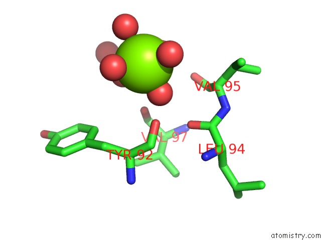
Mono view
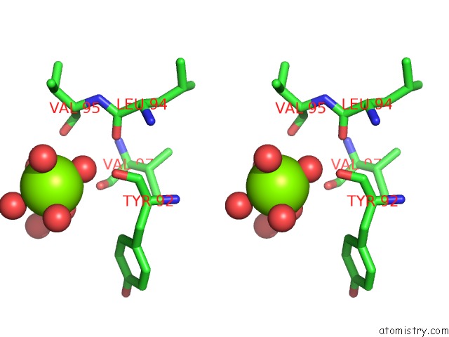
Stereo pair view
Mono view
Stereo pair view
A full contact list of Magnesium with other atoms in the Mg binding
site number 1 of Crystal Structure of Acetolactate Synthase- Small Subunit From Thermotoga Maritima within 5.0Å range:
|
Reference:
J.J.Petkowski,
M.Chruszcz,
M.D.Zimmerman,
H.Zheng,
T.Skarina,
O.Onopriyenko,
M.T.Cymborowski,
K.D.Koclega,
A.Savchenko,
A.Edwards,
W.Minor.
Crystal Structures of TM0549 and NE1324--Two Orthologs of E. Coli Ahas Isozyme III Small Regulatory Subunit. Protein Sci. V. 16 1360 2007.
ISSN: ISSN 0961-8368
PubMed: 17586771
DOI: 10.1110/PS.072793807
Page generated: Tue Aug 13 23:06:46 2024
ISSN: ISSN 0961-8368
PubMed: 17586771
DOI: 10.1110/PS.072793807
Last articles
Zn in 9J0NZn in 9J0O
Zn in 9J0P
Zn in 9FJX
Zn in 9EKB
Zn in 9C0F
Zn in 9CAH
Zn in 9CH0
Zn in 9CH3
Zn in 9CH1