Magnesium »
PDB 2f6y-2fl2 »
2fjm »
Magnesium in PDB 2fjm: The Structure of Phosphotyrosine Phosphatase 1B in Complex with Compound 2
Enzymatic activity of The Structure of Phosphotyrosine Phosphatase 1B in Complex with Compound 2
All present enzymatic activity of The Structure of Phosphotyrosine Phosphatase 1B in Complex with Compound 2:
3.1.3.48;
3.1.3.48;
Protein crystallography data
The structure of The Structure of Phosphotyrosine Phosphatase 1B in Complex with Compound 2, PDB code: 2fjm
was solved by
E.Asante-Appiah,
S.Patel,
C.Desponts,
J.M.Taylor,
C.Lau,
C.Dufresne,
M.Therien,
R.Friesen,
J.W.Becker,
Y.Leblanc,
G.Scapin,
with X-Ray Crystallography technique. A brief refinement statistics is given in the table below:
| Resolution Low / High (Å) | 15.00 / 2.10 |
| Space group | P 21 21 21 |
| Cell size a, b, c (Å), α, β, γ (°) | 86.431, 86.621, 139.193, 90.00, 90.00, 90.00 |
| R / Rfree (%) | 19.2 / 21.7 |
Other elements in 2fjm:
The structure of The Structure of Phosphotyrosine Phosphatase 1B in Complex with Compound 2 also contains other interesting chemical elements:
| Fluorine | (F) | 4 atoms |
| Chlorine | (Cl) | 2 atoms |
Magnesium Binding Sites:
The binding sites of Magnesium atom in the The Structure of Phosphotyrosine Phosphatase 1B in Complex with Compound 2
(pdb code 2fjm). This binding sites where shown within
5.0 Angstroms radius around Magnesium atom.
In total only one binding site of Magnesium was determined in the The Structure of Phosphotyrosine Phosphatase 1B in Complex with Compound 2, PDB code: 2fjm:
In total only one binding site of Magnesium was determined in the The Structure of Phosphotyrosine Phosphatase 1B in Complex with Compound 2, PDB code: 2fjm:
Magnesium binding site 1 out of 1 in 2fjm
Go back to
Magnesium binding site 1 out
of 1 in the The Structure of Phosphotyrosine Phosphatase 1B in Complex with Compound 2
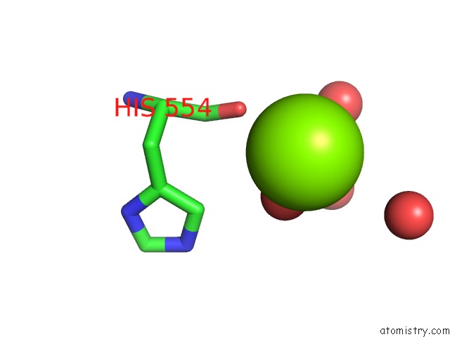
Mono view
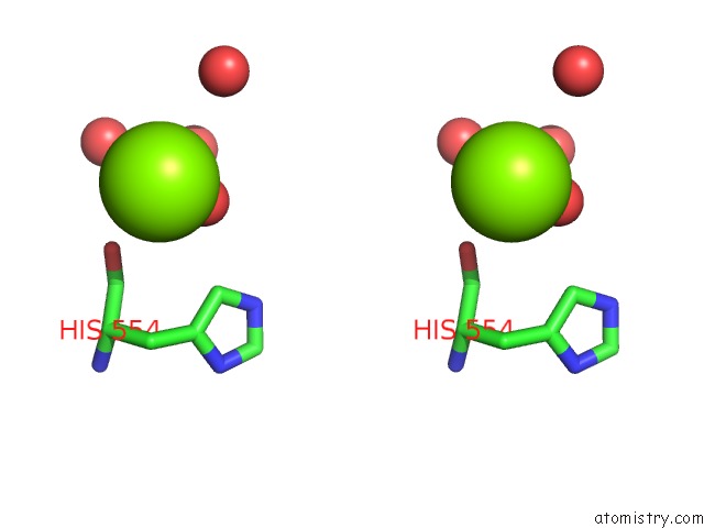
Stereo pair view
Mono view
Stereo pair view
A full contact list of Magnesium with other atoms in the Mg binding
site number 1 of The Structure of Phosphotyrosine Phosphatase 1B in Complex with Compound 2 within 5.0Å range:
|
Reference:
E.Asante-Appiah,
S.Patel,
C.Desponts,
J.M.Taylor,
C.Lau,
C.Dufresne,
M.Therien,
R.Friesen,
J.W.Becker,
Y.Leblanc,
B.P.Kennedy,
G.Scapin.
Conformation-Assisted Inhibition of Protein-Tyrosine Phosphatase-1B Elicits Inhibitor Selectivity Over T-Cell Protein-Tyrosine Phosphatase. J.Biol.Chem. V. 281 8010 2006.
ISSN: ISSN 0021-9258
PubMed: 16407290
DOI: 10.1074/JBC.M511827200
Page generated: Tue Aug 13 23:07:54 2024
ISSN: ISSN 0021-9258
PubMed: 16407290
DOI: 10.1074/JBC.M511827200
Last articles
Zn in 9J0NZn in 9J0O
Zn in 9J0P
Zn in 9FJX
Zn in 9EKB
Zn in 9C0F
Zn in 9CAH
Zn in 9CH0
Zn in 9CH3
Zn in 9CH1