Magnesium »
PDB 2f6y-2fl2 »
2fju »
Magnesium in PDB 2fju: Activated RAC1 Bound to Its Effector Phospholipase C Beta 2
Enzymatic activity of Activated RAC1 Bound to Its Effector Phospholipase C Beta 2
All present enzymatic activity of Activated RAC1 Bound to Its Effector Phospholipase C Beta 2:
3.1.4.11;
3.1.4.11;
Protein crystallography data
The structure of Activated RAC1 Bound to Its Effector Phospholipase C Beta 2, PDB code: 2fju
was solved by
M.R.Jezyk,
J.T.Snyder,
T.K.Harden,
J.Sondek,
with X-Ray Crystallography technique. A brief refinement statistics is given in the table below:
| Resolution Low / High (Å) | 15.00 / 2.20 |
| Space group | P 32 2 1 |
| Cell size a, b, c (Å), α, β, γ (°) | 185.822, 185.822, 93.823, 90.00, 90.00, 120.00 |
| R / Rfree (%) | 20.8 / 22.6 |
Other elements in 2fju:
The structure of Activated RAC1 Bound to Its Effector Phospholipase C Beta 2 also contains other interesting chemical elements:
| Calcium | (Ca) | 1 atom |
Magnesium Binding Sites:
The binding sites of Magnesium atom in the Activated RAC1 Bound to Its Effector Phospholipase C Beta 2
(pdb code 2fju). This binding sites where shown within
5.0 Angstroms radius around Magnesium atom.
In total only one binding site of Magnesium was determined in the Activated RAC1 Bound to Its Effector Phospholipase C Beta 2, PDB code: 2fju:
In total only one binding site of Magnesium was determined in the Activated RAC1 Bound to Its Effector Phospholipase C Beta 2, PDB code: 2fju:
Magnesium binding site 1 out of 1 in 2fju
Go back to
Magnesium binding site 1 out
of 1 in the Activated RAC1 Bound to Its Effector Phospholipase C Beta 2
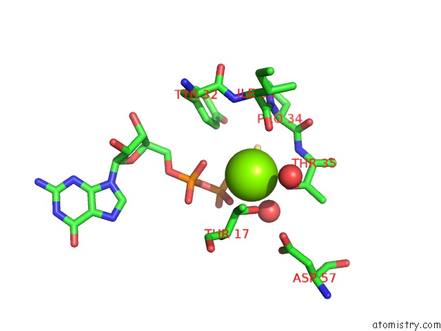
Mono view
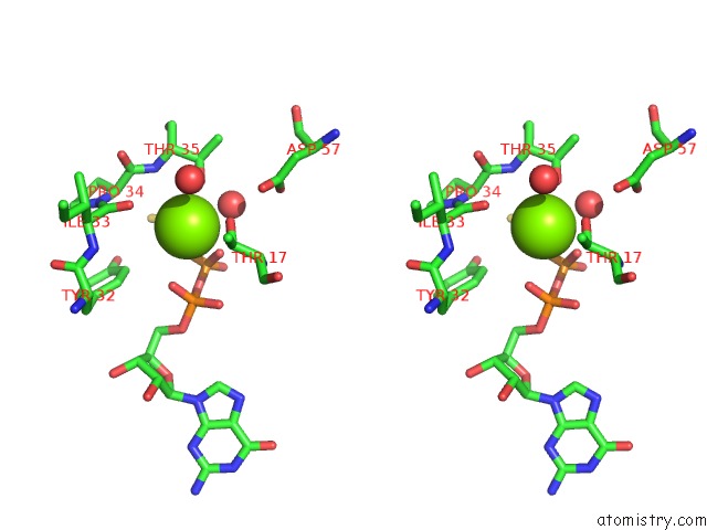
Stereo pair view
Mono view
Stereo pair view
A full contact list of Magnesium with other atoms in the Mg binding
site number 1 of Activated RAC1 Bound to Its Effector Phospholipase C Beta 2 within 5.0Å range:
|
Reference:
M.R.Jezyk,
J.T.Snyder,
S.Gershberg,
D.K.Worthylake,
T.K.Harden,
J.Sondek.
Crystal Structure of RAC1 Bound to Its Effector Phospholipase C-BETA2. Nat.Struct.Mol.Biol. V. 13 1135 2006.
ISSN: ISSN 1545-9993
PubMed: 17115053
DOI: 10.1038/NSMB1175
Page generated: Tue Aug 13 23:07:55 2024
ISSN: ISSN 1545-9993
PubMed: 17115053
DOI: 10.1038/NSMB1175
Last articles
Zn in 9J0NZn in 9J0O
Zn in 9J0P
Zn in 9FJX
Zn in 9EKB
Zn in 9C0F
Zn in 9CAH
Zn in 9CH0
Zn in 9CH3
Zn in 9CH1