Magnesium »
PDB 2f6y-2fl2 »
2fl2 »
Magnesium in PDB 2fl2: Crystal Structure of Ksp in Complex with Inhibitor 19
Protein crystallography data
The structure of Crystal Structure of Ksp in Complex with Inhibitor 19, PDB code: 2fl2
was solved by
Y.Yan,
with X-Ray Crystallography technique. A brief refinement statistics is given in the table below:
| Resolution Low / High (Å) | 50.00 / 2.50 |
| Space group | P 21 21 21 |
| Cell size a, b, c (Å), α, β, γ (°) | 68.800, 79.500, 159.000, 90.00, 90.00, 90.00 |
| R / Rfree (%) | 25.7 / 31.6 |
Other elements in 2fl2:
The structure of Crystal Structure of Ksp in Complex with Inhibitor 19 also contains other interesting chemical elements:
| Fluorine | (F) | 4 atoms |
Magnesium Binding Sites:
The binding sites of Magnesium atom in the Crystal Structure of Ksp in Complex with Inhibitor 19
(pdb code 2fl2). This binding sites where shown within
5.0 Angstroms radius around Magnesium atom.
In total 2 binding sites of Magnesium where determined in the Crystal Structure of Ksp in Complex with Inhibitor 19, PDB code: 2fl2:
Jump to Magnesium binding site number: 1; 2;
In total 2 binding sites of Magnesium where determined in the Crystal Structure of Ksp in Complex with Inhibitor 19, PDB code: 2fl2:
Jump to Magnesium binding site number: 1; 2;
Magnesium binding site 1 out of 2 in 2fl2
Go back to
Magnesium binding site 1 out
of 2 in the Crystal Structure of Ksp in Complex with Inhibitor 19
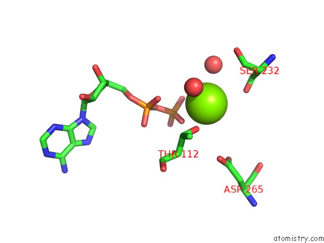
Mono view
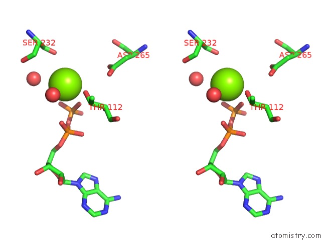
Stereo pair view
Mono view
Stereo pair view
A full contact list of Magnesium with other atoms in the Mg binding
site number 1 of Crystal Structure of Ksp in Complex with Inhibitor 19 within 5.0Å range:
|
Magnesium binding site 2 out of 2 in 2fl2
Go back to
Magnesium binding site 2 out
of 2 in the Crystal Structure of Ksp in Complex with Inhibitor 19
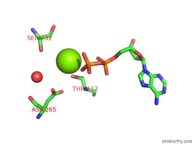
Mono view
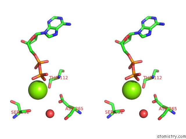
Stereo pair view
Mono view
Stereo pair view
A full contact list of Magnesium with other atoms in the Mg binding
site number 2 of Crystal Structure of Ksp in Complex with Inhibitor 19 within 5.0Å range:
|
Reference:
M.E.Fraley,
R.M.Garbaccio,
K.L.Arrington,
W.F.Hoffman,
E.S.Tasber,
P.J.Coleman,
C.A.Buser,
E.S.Walsh,
K.Hamilton,
C.Fernandes,
M.D.Schaber,
R.B.Lobell,
W.Tao,
V.J.South,
Y.Yan,
L.C.Kuo,
T.Prueksaritanont,
C.Shu,
M.Torrent,
D.C.Heimbrook,
N.E.Kohl,
H.E.Huber,
G.D.Hartman.
Kinesin Spindle Protein (Ksp) Inhibitors. Part 2: the Design, Synthesis, and Characterization of 2,4-Diaryl-2,5-Dihydropyrrole Inhibitors of the Mitotic Kinesin Ksp. Bioorg.Med.Chem.Lett. V. 16 1775 2006.
ISSN: ISSN 0960-894X
PubMed: 16439123
DOI: 10.1016/J.BMCL.2006.01.030
Page generated: Tue Aug 13 23:08:53 2024
ISSN: ISSN 0960-894X
PubMed: 16439123
DOI: 10.1016/J.BMCL.2006.01.030
Last articles
Zn in 9J0NZn in 9J0O
Zn in 9J0P
Zn in 9FJX
Zn in 9EKB
Zn in 9C0F
Zn in 9CAH
Zn in 9CH0
Zn in 9CH3
Zn in 9CH1