Magnesium »
PDB 2fx3-2g9y »
2g07 »
Magnesium in PDB 2g07: X-Ray Structure of Mouse Pyrimidine 5'-Nucleotidase Type 1, Phospho- Enzyme Intermediate Analog with Beryllium Fluoride
Enzymatic activity of X-Ray Structure of Mouse Pyrimidine 5'-Nucleotidase Type 1, Phospho- Enzyme Intermediate Analog with Beryllium Fluoride
All present enzymatic activity of X-Ray Structure of Mouse Pyrimidine 5'-Nucleotidase Type 1, Phospho- Enzyme Intermediate Analog with Beryllium Fluoride:
3.1.1.5;
3.1.1.5;
Protein crystallography data
The structure of X-Ray Structure of Mouse Pyrimidine 5'-Nucleotidase Type 1, Phospho- Enzyme Intermediate Analog with Beryllium Fluoride, PDB code: 2g07
was solved by
E.Bitto,
C.A.Bingman,
G.E.Wesenberg,
G.N.Phillips Jr.,
Center Foreukaryotic Structural Genomics (Cesg),
with X-Ray Crystallography technique. A brief refinement statistics is given in the table below:
| Resolution Low / High (Å) | 67.42 / 2.30 |
| Space group | P 32 |
| Cell size a, b, c (Å), α, β, γ (°) | 134.834, 134.834, 38.945, 90.00, 90.00, 120.00 |
| R / Rfree (%) | 18 / 25.9 |
Other elements in 2g07:
The structure of X-Ray Structure of Mouse Pyrimidine 5'-Nucleotidase Type 1, Phospho- Enzyme Intermediate Analog with Beryllium Fluoride also contains other interesting chemical elements:
| Fluorine | (F) | 6 atoms |
Magnesium Binding Sites:
The binding sites of Magnesium atom in the X-Ray Structure of Mouse Pyrimidine 5'-Nucleotidase Type 1, Phospho- Enzyme Intermediate Analog with Beryllium Fluoride
(pdb code 2g07). This binding sites where shown within
5.0 Angstroms radius around Magnesium atom.
In total 2 binding sites of Magnesium where determined in the X-Ray Structure of Mouse Pyrimidine 5'-Nucleotidase Type 1, Phospho- Enzyme Intermediate Analog with Beryllium Fluoride, PDB code: 2g07:
Jump to Magnesium binding site number: 1; 2;
In total 2 binding sites of Magnesium where determined in the X-Ray Structure of Mouse Pyrimidine 5'-Nucleotidase Type 1, Phospho- Enzyme Intermediate Analog with Beryllium Fluoride, PDB code: 2g07:
Jump to Magnesium binding site number: 1; 2;
Magnesium binding site 1 out of 2 in 2g07
Go back to
Magnesium binding site 1 out
of 2 in the X-Ray Structure of Mouse Pyrimidine 5'-Nucleotidase Type 1, Phospho- Enzyme Intermediate Analog with Beryllium Fluoride
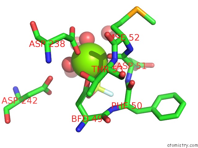
Mono view
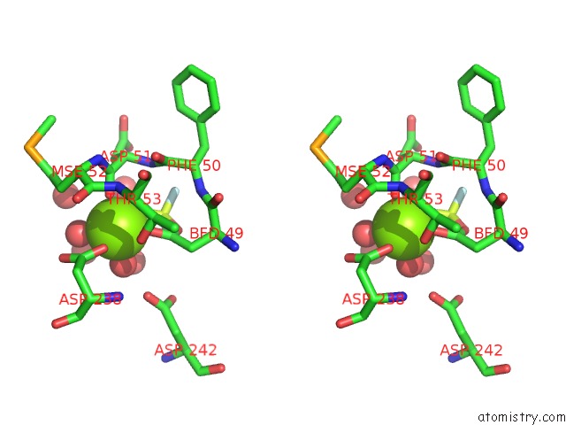
Stereo pair view
Mono view
Stereo pair view
A full contact list of Magnesium with other atoms in the Mg binding
site number 1 of X-Ray Structure of Mouse Pyrimidine 5'-Nucleotidase Type 1, Phospho- Enzyme Intermediate Analog with Beryllium Fluoride within 5.0Å range:
|
Magnesium binding site 2 out of 2 in 2g07
Go back to
Magnesium binding site 2 out
of 2 in the X-Ray Structure of Mouse Pyrimidine 5'-Nucleotidase Type 1, Phospho- Enzyme Intermediate Analog with Beryllium Fluoride
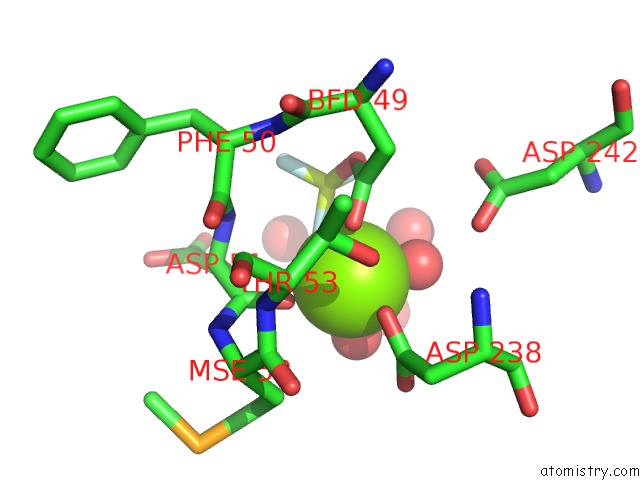
Mono view
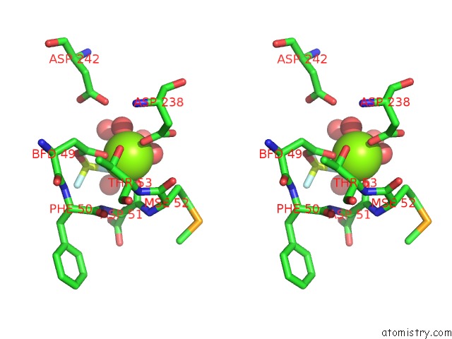
Stereo pair view
Mono view
Stereo pair view
A full contact list of Magnesium with other atoms in the Mg binding
site number 2 of X-Ray Structure of Mouse Pyrimidine 5'-Nucleotidase Type 1, Phospho- Enzyme Intermediate Analog with Beryllium Fluoride within 5.0Å range:
|
Reference:
E.Bitto,
C.A.Bingman,
G.E.Wesenberg,
J.G.Mccoy,
G.N.Phillips.
Structure of Pyrimidine 5'-Nucleotidase Type 1. Insight Into Mechanism of Action and Inhibition During Lead Poisoning. J.Biol.Chem. V. 281 20521 2006.
ISSN: ISSN 0021-9258
PubMed: 16672222
DOI: 10.1074/JBC.M602000200
Page generated: Tue Aug 13 23:20:12 2024
ISSN: ISSN 0021-9258
PubMed: 16672222
DOI: 10.1074/JBC.M602000200
Last articles
Zn in 9J0NZn in 9J0O
Zn in 9J0P
Zn in 9FJX
Zn in 9EKB
Zn in 9C0F
Zn in 9CAH
Zn in 9CH0
Zn in 9CH3
Zn in 9CH1