Magnesium »
PDB 2fx3-2g9y »
2g2s »
Magnesium in PDB 2g2s: Structure of S65G Y66S Gfp Variant After Spontaneous Peptide Hydrolysis
Protein crystallography data
The structure of Structure of S65G Y66S Gfp Variant After Spontaneous Peptide Hydrolysis, PDB code: 2g2s
was solved by
D.P.Barondeau,
with X-Ray Crystallography technique. A brief refinement statistics is given in the table below:
| Resolution Low / High (Å) | 17.80 / 1.20 |
| Space group | P 21 21 21 |
| Cell size a, b, c (Å), α, β, γ (°) | 51.000, 62.300, 68.500, 90.00, 90.00, 90.00 |
| R / Rfree (%) | n/a / 17.4 |
Magnesium Binding Sites:
The binding sites of Magnesium atom in the Structure of S65G Y66S Gfp Variant After Spontaneous Peptide Hydrolysis
(pdb code 2g2s). This binding sites where shown within
5.0 Angstroms radius around Magnesium atom.
In total only one binding site of Magnesium was determined in the Structure of S65G Y66S Gfp Variant After Spontaneous Peptide Hydrolysis, PDB code: 2g2s:
In total only one binding site of Magnesium was determined in the Structure of S65G Y66S Gfp Variant After Spontaneous Peptide Hydrolysis, PDB code: 2g2s:
Magnesium binding site 1 out of 1 in 2g2s
Go back to
Magnesium binding site 1 out
of 1 in the Structure of S65G Y66S Gfp Variant After Spontaneous Peptide Hydrolysis
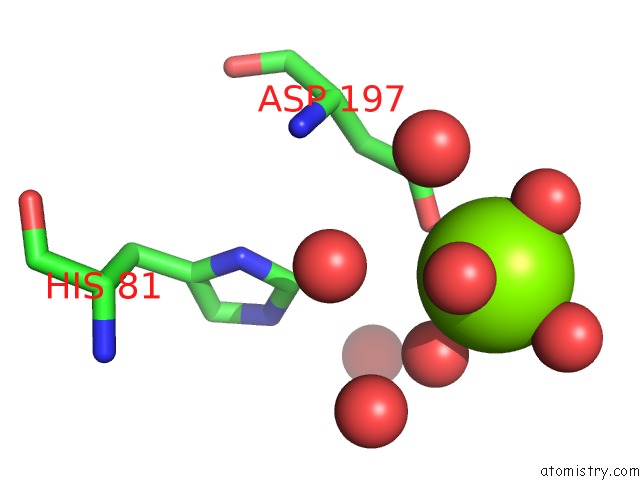
Mono view
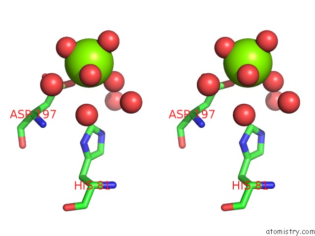
Stereo pair view
Mono view
Stereo pair view
A full contact list of Magnesium with other atoms in the Mg binding
site number 1 of Structure of S65G Y66S Gfp Variant After Spontaneous Peptide Hydrolysis within 5.0Å range:
|
Reference:
D.P.Barondeau,
C.J.Kassmann,
J.A.Tainer,
E.D.Getzoff.
Understanding Gfp Posttranslational Chemistry: Structures of Designed Variants That Achieve Backbone Fragmentation, Hydrolysis, and Decarboxylation. J.Am.Chem.Soc. V. 128 4685 2006.
ISSN: ISSN 0002-7863
PubMed: 16594705
DOI: 10.1021/JA056635L
Page generated: Tue Aug 13 23:22:10 2024
ISSN: ISSN 0002-7863
PubMed: 16594705
DOI: 10.1021/JA056635L
Last articles
Zn in 9J0NZn in 9J0O
Zn in 9J0P
Zn in 9FJX
Zn in 9EKB
Zn in 9C0F
Zn in 9CAH
Zn in 9CH0
Zn in 9CH3
Zn in 9CH1