Magnesium »
PDB 2fx3-2g9y »
2g9y »
Magnesium in PDB 2g9y: Structure of S102T E. Coli Alkaline Phosphatase in Presence of Phosphate at 2.00 A Resolution
Enzymatic activity of Structure of S102T E. Coli Alkaline Phosphatase in Presence of Phosphate at 2.00 A Resolution
All present enzymatic activity of Structure of S102T E. Coli Alkaline Phosphatase in Presence of Phosphate at 2.00 A Resolution:
3.1.3.1;
3.1.3.1;
Protein crystallography data
The structure of Structure of S102T E. Coli Alkaline Phosphatase in Presence of Phosphate at 2.00 A Resolution, PDB code: 2g9y
was solved by
J.Wang,
E.R.Kantrowitz,
with X-Ray Crystallography technique. A brief refinement statistics is given in the table below:
| Resolution Low / High (Å) | 32.70 / 2.00 |
| Space group | I 2 2 2 |
| Cell size a, b, c (Å), α, β, γ (°) | 76.460, 164.490, 192.530, 90.00, 90.00, 90.00 |
| R / Rfree (%) | 20.5 / 24.2 |
Other elements in 2g9y:
The structure of Structure of S102T E. Coli Alkaline Phosphatase in Presence of Phosphate at 2.00 A Resolution also contains other interesting chemical elements:
| Zinc | (Zn) | 4 atoms |
Magnesium Binding Sites:
The binding sites of Magnesium atom in the Structure of S102T E. Coli Alkaline Phosphatase in Presence of Phosphate at 2.00 A Resolution
(pdb code 2g9y). This binding sites where shown within
5.0 Angstroms radius around Magnesium atom.
In total 2 binding sites of Magnesium where determined in the Structure of S102T E. Coli Alkaline Phosphatase in Presence of Phosphate at 2.00 A Resolution, PDB code: 2g9y:
Jump to Magnesium binding site number: 1; 2;
In total 2 binding sites of Magnesium where determined in the Structure of S102T E. Coli Alkaline Phosphatase in Presence of Phosphate at 2.00 A Resolution, PDB code: 2g9y:
Jump to Magnesium binding site number: 1; 2;
Magnesium binding site 1 out of 2 in 2g9y
Go back to
Magnesium binding site 1 out
of 2 in the Structure of S102T E. Coli Alkaline Phosphatase in Presence of Phosphate at 2.00 A Resolution
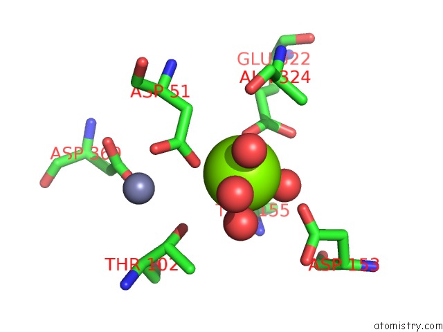
Mono view
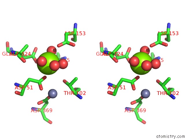
Stereo pair view
Mono view
Stereo pair view
A full contact list of Magnesium with other atoms in the Mg binding
site number 1 of Structure of S102T E. Coli Alkaline Phosphatase in Presence of Phosphate at 2.00 A Resolution within 5.0Å range:
|
Magnesium binding site 2 out of 2 in 2g9y
Go back to
Magnesium binding site 2 out
of 2 in the Structure of S102T E. Coli Alkaline Phosphatase in Presence of Phosphate at 2.00 A Resolution
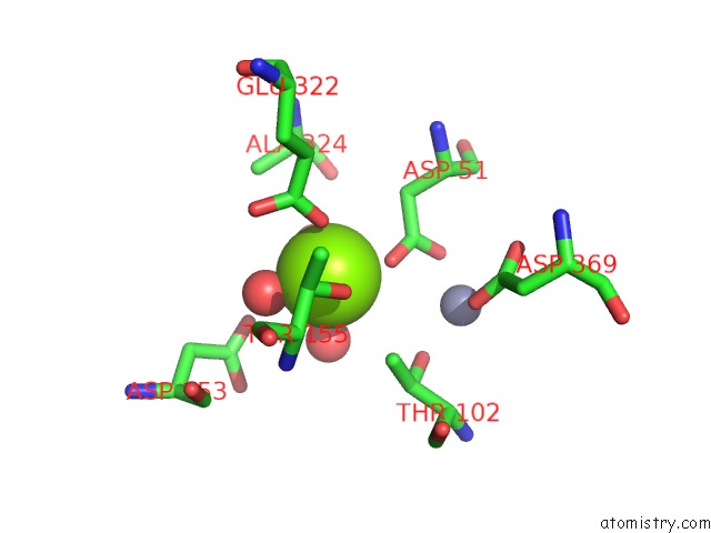
Mono view
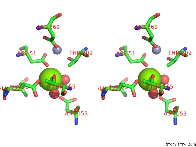
Stereo pair view
Mono view
Stereo pair view
A full contact list of Magnesium with other atoms in the Mg binding
site number 2 of Structure of S102T E. Coli Alkaline Phosphatase in Presence of Phosphate at 2.00 A Resolution within 5.0Å range:
|
Reference:
J.Wang,
E.R.Kantrowitz.
Trapping the Tetrahedral Intermediate in the Alkaline Phosphatase Reaction By Substitution of the Active Site Serine with Threonine. Protein Sci. V. 15 2395 2006.
ISSN: ISSN 0961-8368
PubMed: 17008720
DOI: 10.1110/PS.062351506
Page generated: Tue Aug 13 23:25:10 2024
ISSN: ISSN 0961-8368
PubMed: 17008720
DOI: 10.1110/PS.062351506
Last articles
Zn in 9JYWZn in 9IR4
Zn in 9IR3
Zn in 9GMX
Zn in 9GMW
Zn in 9JEJ
Zn in 9ERF
Zn in 9ERE
Zn in 9EGV
Zn in 9EGW