Magnesium »
PDB 2g9z-2gqp »
2gbl »
Magnesium in PDB 2gbl: Crystal Structure of Full Length Circadian Clock Protein Kaic with Phosphorylation Sites
Enzymatic activity of Crystal Structure of Full Length Circadian Clock Protein Kaic with Phosphorylation Sites
All present enzymatic activity of Crystal Structure of Full Length Circadian Clock Protein Kaic with Phosphorylation Sites:
2.7.1.37;
2.7.1.37;
Protein crystallography data
The structure of Crystal Structure of Full Length Circadian Clock Protein Kaic with Phosphorylation Sites, PDB code: 2gbl
was solved by
R.Pattanayek,
D.R.Williams,
S.Pattanayek,
Y.Xu,
T.Mori,
C.H.Johnson,
P.L.Stewart,
M.Egli,
with X-Ray Crystallography technique. A brief refinement statistics is given in the table below:
| Resolution Low / High (Å) | 30.00 / 2.80 |
| Space group | P 21 21 21 |
| Cell size a, b, c (Å), α, β, γ (°) | 132.873, 135.576, 204.951, 90.00, 90.00, 90.00 |
| R / Rfree (%) | 23 / 29 |
Magnesium Binding Sites:
The binding sites of Magnesium atom in the Crystal Structure of Full Length Circadian Clock Protein Kaic with Phosphorylation Sites
(pdb code 2gbl). This binding sites where shown within
5.0 Angstroms radius around Magnesium atom.
In total 6 binding sites of Magnesium where determined in the Crystal Structure of Full Length Circadian Clock Protein Kaic with Phosphorylation Sites, PDB code: 2gbl:
Jump to Magnesium binding site number: 1; 2; 3; 4; 5; 6;
In total 6 binding sites of Magnesium where determined in the Crystal Structure of Full Length Circadian Clock Protein Kaic with Phosphorylation Sites, PDB code: 2gbl:
Jump to Magnesium binding site number: 1; 2; 3; 4; 5; 6;
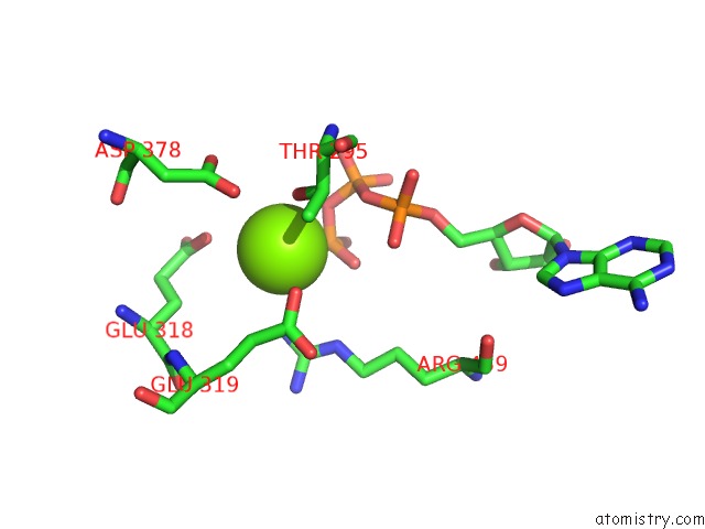
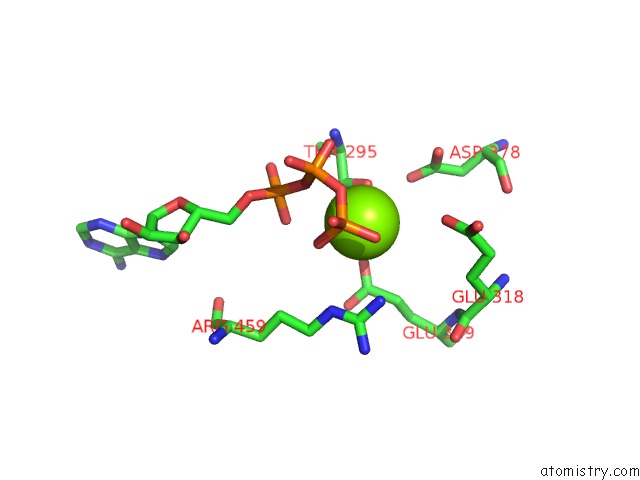
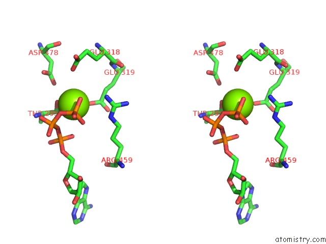
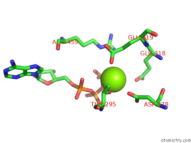
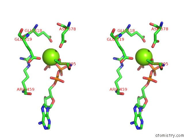
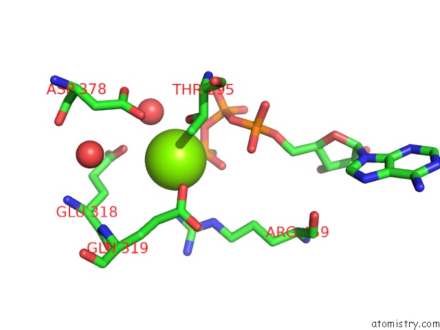
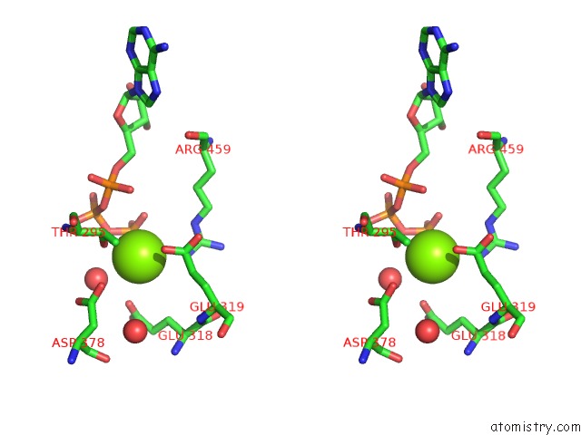
Magnesium binding site 1 out of 6 in 2gbl
Go back to
Magnesium binding site 1 out
of 6 in the Crystal Structure of Full Length Circadian Clock Protein Kaic with Phosphorylation Sites
Mono view
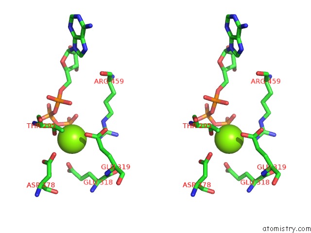
Stereo pair view
Mono view
Stereo pair view
A full contact list of Magnesium with other atoms in the Mg binding
site number 1 of Crystal Structure of Full Length Circadian Clock Protein Kaic with Phosphorylation Sites within 5.0Å range:
|
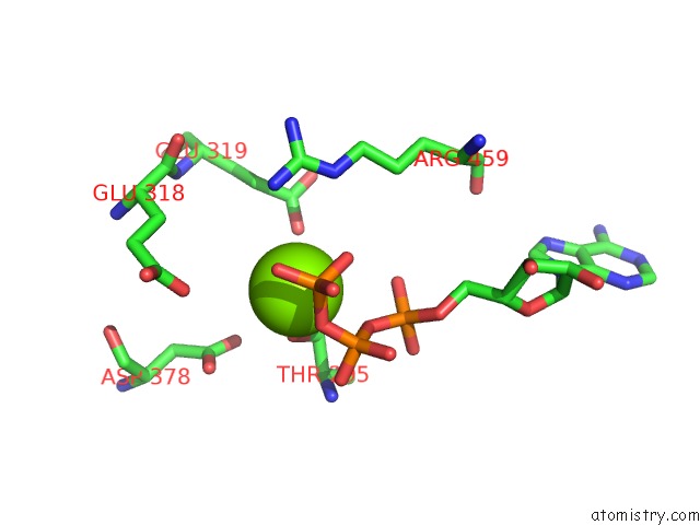
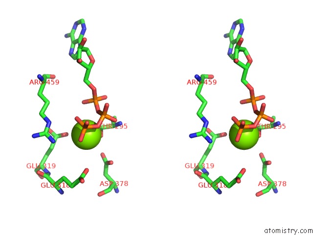
Magnesium binding site 2 out of 6 in 2gbl
Go back to
Magnesium binding site 2 out
of 6 in the Crystal Structure of Full Length Circadian Clock Protein Kaic with Phosphorylation Sites
Mono view
Stereo pair view
Mono view
Stereo pair view
A full contact list of Magnesium with other atoms in the Mg binding
site number 2 of Crystal Structure of Full Length Circadian Clock Protein Kaic with Phosphorylation Sites within 5.0Å range:
|
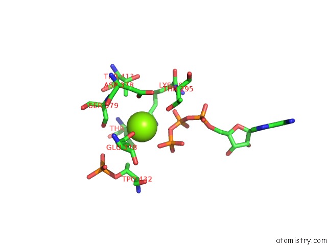
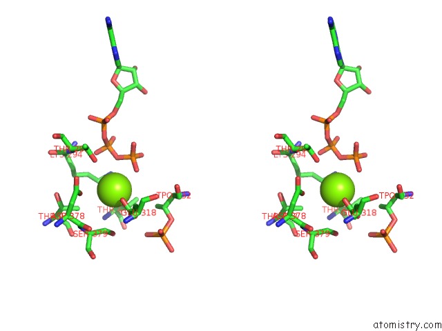
Magnesium binding site 3 out of 6 in 2gbl
Go back to
Magnesium binding site 3 out
of 6 in the Crystal Structure of Full Length Circadian Clock Protein Kaic with Phosphorylation Sites
Mono view
Stereo pair view
Mono view
Stereo pair view
A full contact list of Magnesium with other atoms in the Mg binding
site number 3 of Crystal Structure of Full Length Circadian Clock Protein Kaic with Phosphorylation Sites within 5.0Å range:
|
Magnesium binding site 4 out of 6 in 2gbl
Go back to
Magnesium binding site 4 out
of 6 in the Crystal Structure of Full Length Circadian Clock Protein Kaic with Phosphorylation Sites
Mono view
Stereo pair view
Mono view
Stereo pair view
A full contact list of Magnesium with other atoms in the Mg binding
site number 4 of Crystal Structure of Full Length Circadian Clock Protein Kaic with Phosphorylation Sites within 5.0Å range:
|
Magnesium binding site 5 out of 6 in 2gbl
Go back to
Magnesium binding site 5 out
of 6 in the Crystal Structure of Full Length Circadian Clock Protein Kaic with Phosphorylation Sites
Mono view
Stereo pair view
Mono view
Stereo pair view
A full contact list of Magnesium with other atoms in the Mg binding
site number 5 of Crystal Structure of Full Length Circadian Clock Protein Kaic with Phosphorylation Sites within 5.0Å range:
|
Magnesium binding site 6 out of 6 in 2gbl
Go back to
Magnesium binding site 6 out
of 6 in the Crystal Structure of Full Length Circadian Clock Protein Kaic with Phosphorylation Sites
Mono view
Stereo pair view
Mono view
Stereo pair view
A full contact list of Magnesium with other atoms in the Mg binding
site number 6 of Crystal Structure of Full Length Circadian Clock Protein Kaic with Phosphorylation Sites within 5.0Å range:
|
Reference:
R.Pattanayek,
D.R.Williams,
S.Pattanayek,
Y.Xu,
T.Mori,
C.H.Johnson,
P.L.Stewart,
M.Egli.
Analysis of Kaia-Kaic Protein Interactions in the Cyano-Bacterial Circadian Clock Using Hybrid Structural Methods. Embo J. V. 25 2017 2006.
ISSN: ISSN 0261-4189
PubMed: 16628225
DOI: 10.1038/SJ.EMBOJ.7601086
Page generated: Tue Aug 13 23:25:56 2024
ISSN: ISSN 0261-4189
PubMed: 16628225
DOI: 10.1038/SJ.EMBOJ.7601086
Last articles
Zn in 9JYWZn in 9IR4
Zn in 9IR3
Zn in 9GMX
Zn in 9GMW
Zn in 9JEJ
Zn in 9ERF
Zn in 9ERE
Zn in 9EGV
Zn in 9EGW