Magnesium »
PDB 2gqr-2haw »
2h1c »
Magnesium in PDB 2h1c: Crystal Structure of Fitacb From Neisseria Gonorrhoeae
Protein crystallography data
The structure of Crystal Structure of Fitacb From Neisseria Gonorrhoeae, PDB code: 2h1c
was solved by
K.Mattison,
J.S.Wilbur,
M.So,
R.G.Brennan,
with X-Ray Crystallography technique. A brief refinement statistics is given in the table below:
| Resolution Low / High (Å) | 39.11 / 1.80 |
| Space group | C 1 2 1 |
| Cell size a, b, c (Å), α, β, γ (°) | 69.970, 50.700, 48.280, 90.00, 118.55, 90.00 |
| R / Rfree (%) | 19.1 / 22.3 |
Magnesium Binding Sites:
The binding sites of Magnesium atom in the Crystal Structure of Fitacb From Neisseria Gonorrhoeae
(pdb code 2h1c). This binding sites where shown within
5.0 Angstroms radius around Magnesium atom.
In total 3 binding sites of Magnesium where determined in the Crystal Structure of Fitacb From Neisseria Gonorrhoeae, PDB code: 2h1c:
Jump to Magnesium binding site number: 1; 2; 3;
In total 3 binding sites of Magnesium where determined in the Crystal Structure of Fitacb From Neisseria Gonorrhoeae, PDB code: 2h1c:
Jump to Magnesium binding site number: 1; 2; 3;
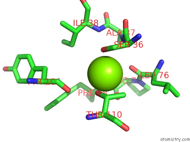
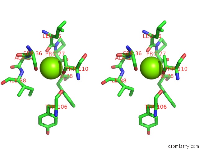
Magnesium binding site 1 out of 3 in 2h1c
Go back to
Magnesium binding site 1 out
of 3 in the Crystal Structure of Fitacb From Neisseria Gonorrhoeae
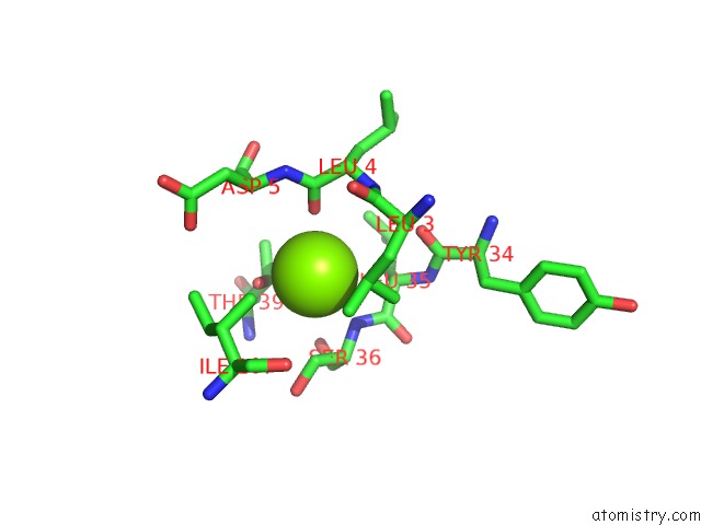
Mono view
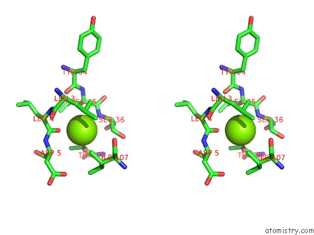
Stereo pair view
Mono view
Stereo pair view
A full contact list of Magnesium with other atoms in the Mg binding
site number 1 of Crystal Structure of Fitacb From Neisseria Gonorrhoeae within 5.0Å range:
|
Magnesium binding site 2 out of 3 in 2h1c
Go back to
Magnesium binding site 2 out
of 3 in the Crystal Structure of Fitacb From Neisseria Gonorrhoeae
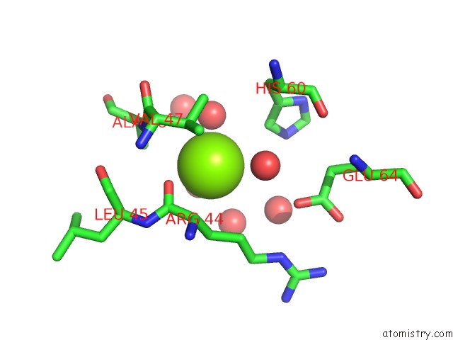
Mono view
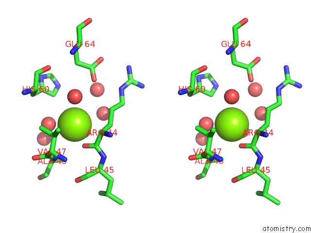
Stereo pair view
Mono view
Stereo pair view
A full contact list of Magnesium with other atoms in the Mg binding
site number 2 of Crystal Structure of Fitacb From Neisseria Gonorrhoeae within 5.0Å range:
|
Magnesium binding site 3 out of 3 in 2h1c
Go back to
Magnesium binding site 3 out
of 3 in the Crystal Structure of Fitacb From Neisseria Gonorrhoeae
Mono view
Stereo pair view
Mono view
Stereo pair view
A full contact list of Magnesium with other atoms in the Mg binding
site number 3 of Crystal Structure of Fitacb From Neisseria Gonorrhoeae within 5.0Å range:
|
Reference:
K.Mattison,
J.S.Wilbur,
M.So,
R.G.Brennan.
Structure of Fitab From Neisseria Gonorrhoeae Bound to Dna Reveals A Tetramer of Toxin-Antitoxin Heterodimers Containing Pin Domains and Ribbon-Helix-Helix Motifs. J.Biol.Chem. V. 281 37942 2006.
ISSN: ISSN 0021-9258
PubMed: 16982615
DOI: 10.1074/JBC.M605198200
Page generated: Tue Aug 13 23:40:41 2024
ISSN: ISSN 0021-9258
PubMed: 16982615
DOI: 10.1074/JBC.M605198200
Last articles
Fe in 2YXOFe in 2YRS
Fe in 2YXC
Fe in 2YNM
Fe in 2YVJ
Fe in 2YP1
Fe in 2YU2
Fe in 2YU1
Fe in 2YQB
Fe in 2YOO