Magnesium »
PDB 2gqr-2haw »
2h7v »
Magnesium in PDB 2h7v: Co-Crystal Structure of Ypka-RAC1
Protein crystallography data
The structure of Co-Crystal Structure of Ypka-RAC1, PDB code: 2h7v
was solved by
G.Prehna,
M.Ivanov,
J.B.Bliska,
C.E.Stebbins,
with X-Ray Crystallography technique. A brief refinement statistics is given in the table below:
| Resolution Low / High (Å) | 95.78 / 2.60 |
| Space group | P 1 |
| Cell size a, b, c (Å), α, β, γ (°) | 66.398, 75.519, 99.772, 92.08, 103.38, 115.79 |
| R / Rfree (%) | 22.2 / 25.7 |
Magnesium Binding Sites:
The binding sites of Magnesium atom in the Co-Crystal Structure of Ypka-RAC1
(pdb code 2h7v). This binding sites where shown within
5.0 Angstroms radius around Magnesium atom.
In total 2 binding sites of Magnesium where determined in the Co-Crystal Structure of Ypka-RAC1, PDB code: 2h7v:
Jump to Magnesium binding site number: 1; 2;
In total 2 binding sites of Magnesium where determined in the Co-Crystal Structure of Ypka-RAC1, PDB code: 2h7v:
Jump to Magnesium binding site number: 1; 2;
Magnesium binding site 1 out of 2 in 2h7v
Go back to
Magnesium binding site 1 out
of 2 in the Co-Crystal Structure of Ypka-RAC1
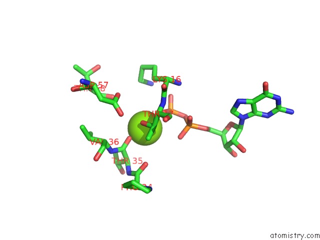
Mono view
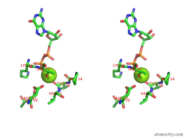
Stereo pair view
Mono view
Stereo pair view
A full contact list of Magnesium with other atoms in the Mg binding
site number 1 of Co-Crystal Structure of Ypka-RAC1 within 5.0Å range:
|
Magnesium binding site 2 out of 2 in 2h7v
Go back to
Magnesium binding site 2 out
of 2 in the Co-Crystal Structure of Ypka-RAC1
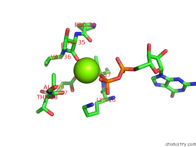
Mono view
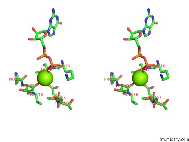
Stereo pair view
Mono view
Stereo pair view
A full contact list of Magnesium with other atoms in the Mg binding
site number 2 of Co-Crystal Structure of Ypka-RAC1 within 5.0Å range:
|
Reference:
G.Prehna,
M.I.Ivanov,
J.B.Bliska,
C.E.Stebbins.
Yersinia Virulence Depends on Mimicry of Host Rho-Family Nucleotide Dissociation Inhibitors. Cell(Cambridge,Mass.) V. 126 869 2006.
ISSN: ISSN 0092-8674
PubMed: 16959567
DOI: 10.1016/J.CELL.2006.06.056
Page generated: Tue Aug 13 23:43:32 2024
ISSN: ISSN 0092-8674
PubMed: 16959567
DOI: 10.1016/J.CELL.2006.06.056
Last articles
Zn in 9MJ5Zn in 9HNW
Zn in 9G0L
Zn in 9FNE
Zn in 9DZN
Zn in 9E0I
Zn in 9D32
Zn in 9DAK
Zn in 8ZXC
Zn in 8ZUF