Magnesium »
PDB 2hb4-2hkj »
2hbv »
Magnesium in PDB 2hbv: Crystal Structure of Alpha-Amino-Beta-Carboxymuconate-Epsilon- Semialdehyde-Decarboxylase (Acmsd)
Enzymatic activity of Crystal Structure of Alpha-Amino-Beta-Carboxymuconate-Epsilon- Semialdehyde-Decarboxylase (Acmsd)
All present enzymatic activity of Crystal Structure of Alpha-Amino-Beta-Carboxymuconate-Epsilon- Semialdehyde-Decarboxylase (Acmsd):
4.1.1.45;
4.1.1.45;
Protein crystallography data
The structure of Crystal Structure of Alpha-Amino-Beta-Carboxymuconate-Epsilon- Semialdehyde-Decarboxylase (Acmsd), PDB code: 2hbv
was solved by
D.Martynowski,
Y.Eyobo,
T.Li,
K.Yang,
A.Liu,
H.Zhang,
with X-Ray Crystallography technique. A brief refinement statistics is given in the table below:
| Resolution Low / High (Å) | 33.81 / 1.65 |
| Space group | C 1 2 1 |
| Cell size a, b, c (Å), α, β, γ (°) | 153.557, 48.107, 110.700, 90.00, 127.31, 90.00 |
| R / Rfree (%) | 21.1 / 25.1 |
Other elements in 2hbv:
The structure of Crystal Structure of Alpha-Amino-Beta-Carboxymuconate-Epsilon- Semialdehyde-Decarboxylase (Acmsd) also contains other interesting chemical elements:
| Zinc | (Zn) | 2 atoms |
Magnesium Binding Sites:
The binding sites of Magnesium atom in the Crystal Structure of Alpha-Amino-Beta-Carboxymuconate-Epsilon- Semialdehyde-Decarboxylase (Acmsd)
(pdb code 2hbv). This binding sites where shown within
5.0 Angstroms radius around Magnesium atom.
In total only one binding site of Magnesium was determined in the Crystal Structure of Alpha-Amino-Beta-Carboxymuconate-Epsilon- Semialdehyde-Decarboxylase (Acmsd), PDB code: 2hbv:
In total only one binding site of Magnesium was determined in the Crystal Structure of Alpha-Amino-Beta-Carboxymuconate-Epsilon- Semialdehyde-Decarboxylase (Acmsd), PDB code: 2hbv:
Magnesium binding site 1 out of 1 in 2hbv
Go back to
Magnesium binding site 1 out
of 1 in the Crystal Structure of Alpha-Amino-Beta-Carboxymuconate-Epsilon- Semialdehyde-Decarboxylase (Acmsd)
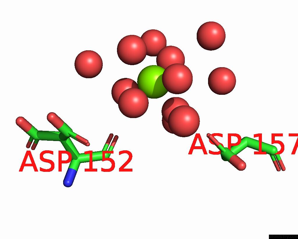
Mono view
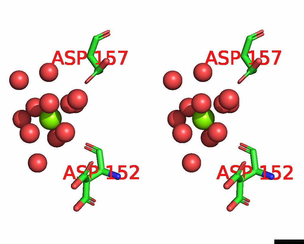
Stereo pair view
Mono view
Stereo pair view
A full contact list of Magnesium with other atoms in the Mg binding
site number 1 of Crystal Structure of Alpha-Amino-Beta-Carboxymuconate-Epsilon- Semialdehyde-Decarboxylase (Acmsd) within 5.0Å range:
|
Reference:
D.Martynowski,
Y.Eyobo,
T.Li,
K.Yang,
A.Liu,
H.Zhang.
Crystal Structure of Alpha-Amino-Beta-Carboxymuconate-Epsilon-Semialdehyde Decarboxylase: Insight Into the Active Site and Catalytic Mechanism of A Novel Decarboxylation Reaction. Biochemistry V. 45 10412 2006.
ISSN: ISSN 0006-2960
PubMed: 16939194
DOI: 10.1021/BI060903Q
Page generated: Tue Aug 13 23:48:55 2024
ISSN: ISSN 0006-2960
PubMed: 16939194
DOI: 10.1021/BI060903Q
Last articles
Zn in 9JYWZn in 9IR4
Zn in 9IR3
Zn in 9GMX
Zn in 9GMW
Zn in 9JEJ
Zn in 9ERF
Zn in 9ERE
Zn in 9EGV
Zn in 9EGW