Magnesium »
PDB 2hb4-2hkj »
2hfj »
Magnesium in PDB 2hfj: Pikromycin Thioesterase with Covalent Pentaketide Affinity Label
Protein crystallography data
The structure of Pikromycin Thioesterase with Covalent Pentaketide Affinity Label, PDB code: 2hfj
was solved by
D.L.Akey,
J.D.Kittendorf,
J.W.Giraldes,
R.A.Fecik,
D.H.Sherman,
J.L.Smith,
with X-Ray Crystallography technique. A brief refinement statistics is given in the table below:
| Resolution Low / High (Å) | 50.00 / 1.95 |
| Space group | P 21 21 2 |
| Cell size a, b, c (Å), α, β, γ (°) | 107.686, 131.063, 56.864, 90.00, 90.00, 90.00 |
| R / Rfree (%) | 20 / 24 |
Magnesium Binding Sites:
The binding sites of Magnesium atom in the Pikromycin Thioesterase with Covalent Pentaketide Affinity Label
(pdb code 2hfj). This binding sites where shown within
5.0 Angstroms radius around Magnesium atom.
In total only one binding site of Magnesium was determined in the Pikromycin Thioesterase with Covalent Pentaketide Affinity Label, PDB code: 2hfj:
In total only one binding site of Magnesium was determined in the Pikromycin Thioesterase with Covalent Pentaketide Affinity Label, PDB code: 2hfj:
Magnesium binding site 1 out of 1 in 2hfj
Go back to
Magnesium binding site 1 out
of 1 in the Pikromycin Thioesterase with Covalent Pentaketide Affinity Label
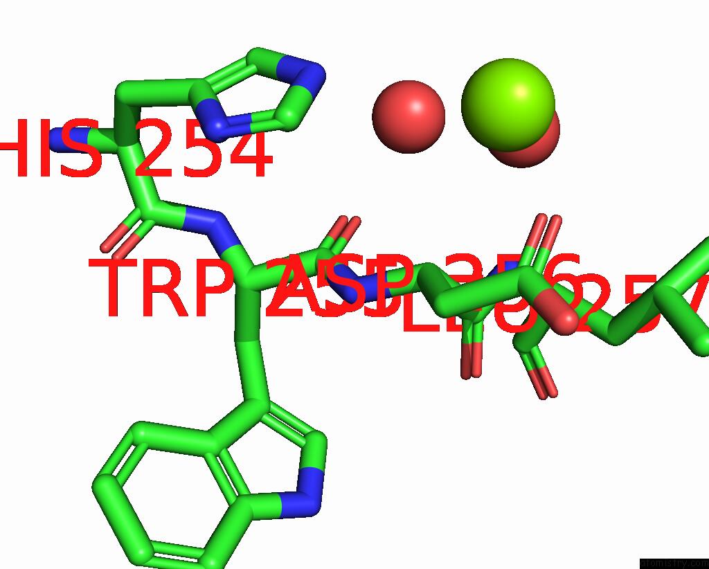
Mono view
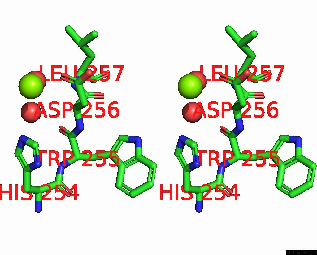
Stereo pair view
Mono view
Stereo pair view
A full contact list of Magnesium with other atoms in the Mg binding
site number 1 of Pikromycin Thioesterase with Covalent Pentaketide Affinity Label within 5.0Å range:
|
Reference:
D.L.Akey,
J.D.Kittendorf,
J.W.Giraldes,
R.A.Fecik,
D.H.Sherman,
J.L.Smith.
Structural Basis For Macrolactonization By the Pikromycin Thioesterase Nat.Chem.Biol. V. 2 537 2006.
ISSN: ISSN 1552-4450
PubMed: 16969372
DOI: 10.1038/NCHEMBIO824
Page generated: Tue Aug 13 23:51:01 2024
ISSN: ISSN 1552-4450
PubMed: 16969372
DOI: 10.1038/NCHEMBIO824
Last articles
Zn in 9J0NZn in 9J0O
Zn in 9J0P
Zn in 9FJX
Zn in 9EKB
Zn in 9C0F
Zn in 9CAH
Zn in 9CH0
Zn in 9CH3
Zn in 9CH1