Magnesium »
PDB 2hb4-2hkj »
2hhq »
Magnesium in PDB 2hhq: O6-Methyl-Guanine:T Pair in the Polymerase-10 Basepair Position
Enzymatic activity of O6-Methyl-Guanine:T Pair in the Polymerase-10 Basepair Position
All present enzymatic activity of O6-Methyl-Guanine:T Pair in the Polymerase-10 Basepair Position:
2.7.7.7;
2.7.7.7;
Protein crystallography data
The structure of O6-Methyl-Guanine:T Pair in the Polymerase-10 Basepair Position, PDB code: 2hhq
was solved by
J.J.Warren,
L.J.Forsberg,
L.S.Beese,
with X-Ray Crystallography technique. A brief refinement statistics is given in the table below:
| Resolution Low / High (Å) | 50.00 / 1.80 |
| Space group | P 21 21 21 |
| Cell size a, b, c (Å), α, β, γ (°) | 86.792, 93.274, 106.018, 90.00, 90.00, 90.00 |
| R / Rfree (%) | 21.2 / 24.4 |
Magnesium Binding Sites:
The binding sites of Magnesium atom in the O6-Methyl-Guanine:T Pair in the Polymerase-10 Basepair Position
(pdb code 2hhq). This binding sites where shown within
5.0 Angstroms radius around Magnesium atom.
In total only one binding site of Magnesium was determined in the O6-Methyl-Guanine:T Pair in the Polymerase-10 Basepair Position, PDB code: 2hhq:
In total only one binding site of Magnesium was determined in the O6-Methyl-Guanine:T Pair in the Polymerase-10 Basepair Position, PDB code: 2hhq:
Magnesium binding site 1 out of 1 in 2hhq
Go back to
Magnesium binding site 1 out
of 1 in the O6-Methyl-Guanine:T Pair in the Polymerase-10 Basepair Position
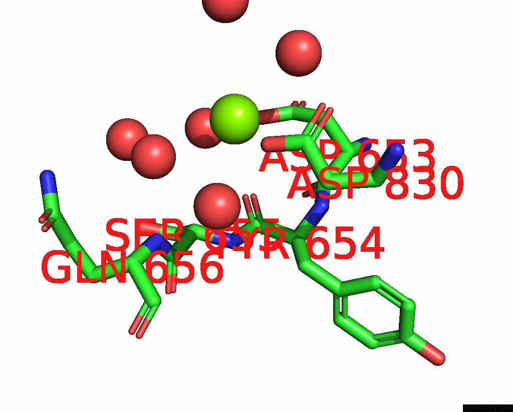
Mono view
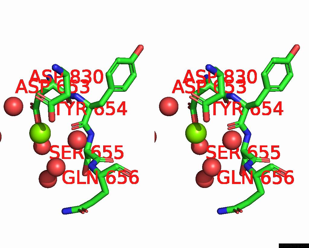
Stereo pair view
Mono view
Stereo pair view
A full contact list of Magnesium with other atoms in the Mg binding
site number 1 of O6-Methyl-Guanine:T Pair in the Polymerase-10 Basepair Position within 5.0Å range:
|
Reference:
J.J.Warren,
L.J.Forsberg,
L.S.Beese.
The Structural Basis For the Mutagenicity of O6-Methyl-Guanine Lesions. Proc.Natl.Acad.Sci.Usa V. 103 19701 2006.
ISSN: ISSN 0027-8424
PubMed: 17179038
DOI: 10.1073/PNAS.0609580103
Page generated: Tue Aug 13 23:53:01 2024
ISSN: ISSN 0027-8424
PubMed: 17179038
DOI: 10.1073/PNAS.0609580103
Last articles
Cl in 8DCQCl in 8DCG
Cl in 8DB5
Cl in 8DCO
Cl in 8DCH
Cl in 8DCF
Cl in 8DCD
Cl in 8DC9
Cl in 8DAK
Cl in 8DCB