Magnesium »
PDB 2hld-2hxf »
2hne »
Magnesium in PDB 2hne: Crystal Structure of L-Fuconate Dehydratase From Xanthomonas Campestris Pv. Campestris Str. Atcc 33913
Enzymatic activity of Crystal Structure of L-Fuconate Dehydratase From Xanthomonas Campestris Pv. Campestris Str. Atcc 33913
All present enzymatic activity of Crystal Structure of L-Fuconate Dehydratase From Xanthomonas Campestris Pv. Campestris Str. Atcc 33913:
4.2.1.68;
4.2.1.68;
Protein crystallography data
The structure of Crystal Structure of L-Fuconate Dehydratase From Xanthomonas Campestris Pv. Campestris Str. Atcc 33913, PDB code: 2hne
was solved by
A.A.Fedorov,
E.V.Fedorov,
W.S.Yew,
J.A.Gerlt,
S.C.Almo,
S.K.Burley,
Newyork Sgx Research Center For Structural Genomics (Nysgxrc),
with X-Ray Crystallography technique. A brief refinement statistics is given in the table below:
| Resolution Low / High (Å) | 24.68 / 2.00 |
| Space group | P 32 2 1 |
| Cell size a, b, c (Å), α, β, γ (°) | 130.633, 130.633, 195.829, 90.00, 90.00, 120.00 |
| R / Rfree (%) | 24.1 / 27.4 |
Magnesium Binding Sites:
The binding sites of Magnesium atom in the Crystal Structure of L-Fuconate Dehydratase From Xanthomonas Campestris Pv. Campestris Str. Atcc 33913
(pdb code 2hne). This binding sites where shown within
5.0 Angstroms radius around Magnesium atom.
In total 4 binding sites of Magnesium where determined in the Crystal Structure of L-Fuconate Dehydratase From Xanthomonas Campestris Pv. Campestris Str. Atcc 33913, PDB code: 2hne:
Jump to Magnesium binding site number: 1; 2; 3; 4;
In total 4 binding sites of Magnesium where determined in the Crystal Structure of L-Fuconate Dehydratase From Xanthomonas Campestris Pv. Campestris Str. Atcc 33913, PDB code: 2hne:
Jump to Magnesium binding site number: 1; 2; 3; 4;
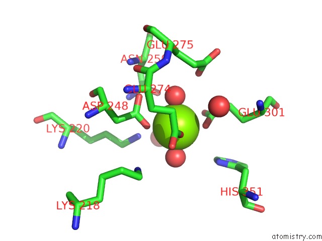
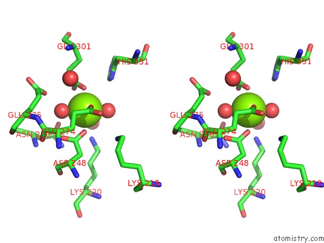
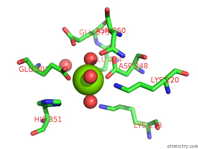
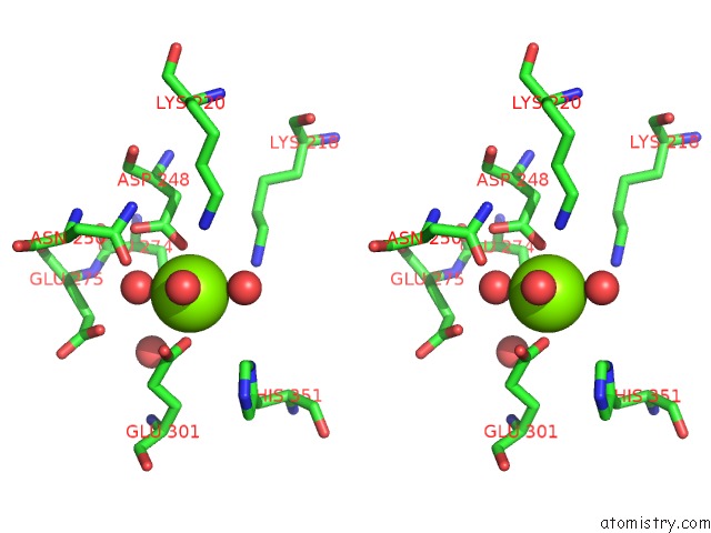
Magnesium binding site 1 out of 4 in 2hne
Go back to
Magnesium binding site 1 out
of 4 in the Crystal Structure of L-Fuconate Dehydratase From Xanthomonas Campestris Pv. Campestris Str. Atcc 33913
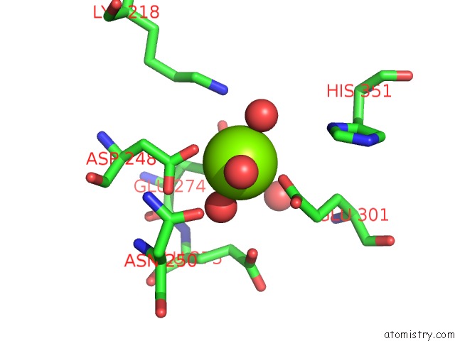
Mono view
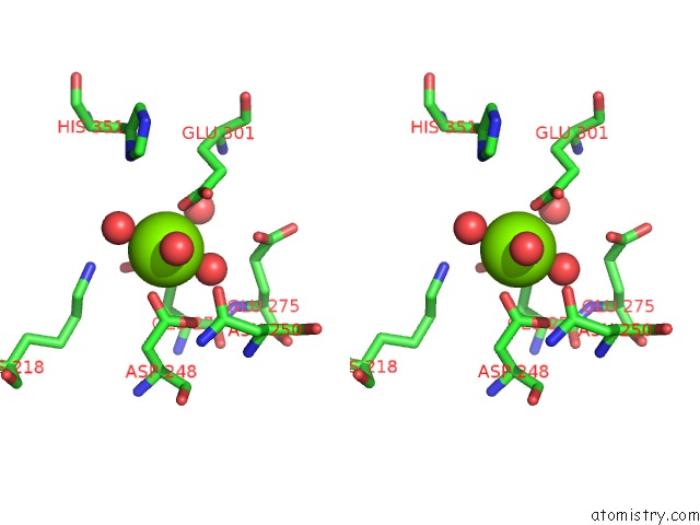
Stereo pair view
Mono view
Stereo pair view
A full contact list of Magnesium with other atoms in the Mg binding
site number 1 of Crystal Structure of L-Fuconate Dehydratase From Xanthomonas Campestris Pv. Campestris Str. Atcc 33913 within 5.0Å range:
|
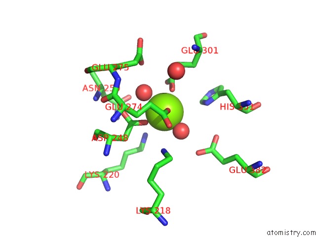
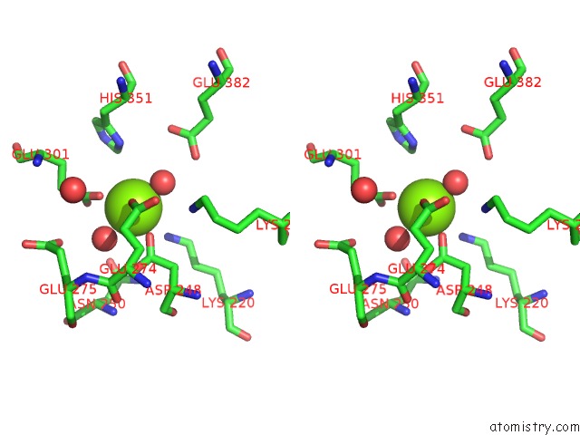
Magnesium binding site 2 out of 4 in 2hne
Go back to
Magnesium binding site 2 out
of 4 in the Crystal Structure of L-Fuconate Dehydratase From Xanthomonas Campestris Pv. Campestris Str. Atcc 33913
Mono view
Stereo pair view
Mono view
Stereo pair view
A full contact list of Magnesium with other atoms in the Mg binding
site number 2 of Crystal Structure of L-Fuconate Dehydratase From Xanthomonas Campestris Pv. Campestris Str. Atcc 33913 within 5.0Å range:
|
Magnesium binding site 3 out of 4 in 2hne
Go back to
Magnesium binding site 3 out
of 4 in the Crystal Structure of L-Fuconate Dehydratase From Xanthomonas Campestris Pv. Campestris Str. Atcc 33913
Mono view
Stereo pair view
Mono view
Stereo pair view
A full contact list of Magnesium with other atoms in the Mg binding
site number 3 of Crystal Structure of L-Fuconate Dehydratase From Xanthomonas Campestris Pv. Campestris Str. Atcc 33913 within 5.0Å range:
|
Magnesium binding site 4 out of 4 in 2hne
Go back to
Magnesium binding site 4 out
of 4 in the Crystal Structure of L-Fuconate Dehydratase From Xanthomonas Campestris Pv. Campestris Str. Atcc 33913
Mono view
Stereo pair view
Mono view
Stereo pair view
A full contact list of Magnesium with other atoms in the Mg binding
site number 4 of Crystal Structure of L-Fuconate Dehydratase From Xanthomonas Campestris Pv. Campestris Str. Atcc 33913 within 5.0Å range:
|
Reference:
A.A.Fedorov,
E.V.Fedorov,
W.S.Yew,
J.A.Gerlt,
S.C.Almo.
Crystal Structure of L-Fuconate Dehydratase From Xanthomonas Campestris Pv. Campestris Str. Atcc 33913 To Be Published.
Page generated: Sun Aug 10 11:24:50 2025
Last articles
Mg in 6I0SMg in 6HZM
Mg in 6I03
Mg in 6HZ7
Mg in 6HZ6
Mg in 6HZ5
Mg in 6HXH
Mg in 6HZ4
Mg in 6HYS
Mg in 6HYT