Magnesium »
PDB 2hld-2hxf »
2hom »
Magnesium in PDB 2hom: Crystal Structure of An E. Coli Thi-Box Riboswitch Bound to Thiamine Monophosphate
Protein crystallography data
The structure of Crystal Structure of An E. Coli Thi-Box Riboswitch Bound to Thiamine Monophosphate, PDB code: 2hom
was solved by
T.E.Edwards,
A.R.Ferre-D'amare,
with X-Ray Crystallography technique. A brief refinement statistics is given in the table below:
| Resolution Low / High (Å) | 30.00 / 2.89 |
| Space group | P 32 1 2 |
| Cell size a, b, c (Å), α, β, γ (°) | 65.700, 65.700, 101.700, 90.00, 90.00, 120.00 |
| R / Rfree (%) | 20.3 / 26.6 |
Other elements in 2hom:
The structure of Crystal Structure of An E. Coli Thi-Box Riboswitch Bound to Thiamine Monophosphate also contains other interesting chemical elements:
| Calcium | (Ca) | 2 atoms |
Magnesium Binding Sites:
The binding sites of Magnesium atom in the Crystal Structure of An E. Coli Thi-Box Riboswitch Bound to Thiamine Monophosphate
(pdb code 2hom). This binding sites where shown within
5.0 Angstroms radius around Magnesium atom.
In total 4 binding sites of Magnesium where determined in the Crystal Structure of An E. Coli Thi-Box Riboswitch Bound to Thiamine Monophosphate, PDB code: 2hom:
Jump to Magnesium binding site number: 1; 2; 3; 4;
In total 4 binding sites of Magnesium where determined in the Crystal Structure of An E. Coli Thi-Box Riboswitch Bound to Thiamine Monophosphate, PDB code: 2hom:
Jump to Magnesium binding site number: 1; 2; 3; 4;
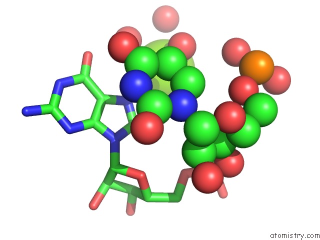
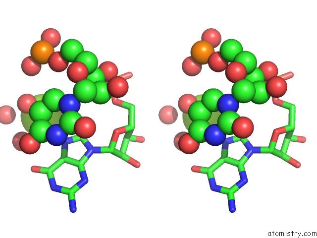
Magnesium binding site 1 out of 4 in 2hom
Go back to
Magnesium binding site 1 out
of 4 in the Crystal Structure of An E. Coli Thi-Box Riboswitch Bound to Thiamine Monophosphate
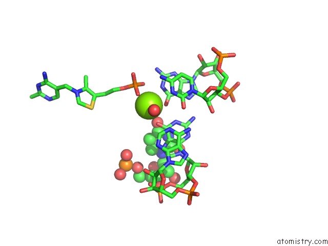
Mono view
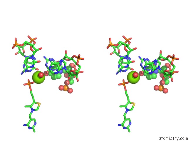
Stereo pair view
Mono view
Stereo pair view
A full contact list of Magnesium with other atoms in the Mg binding
site number 1 of Crystal Structure of An E. Coli Thi-Box Riboswitch Bound to Thiamine Monophosphate within 5.0Å range:
|
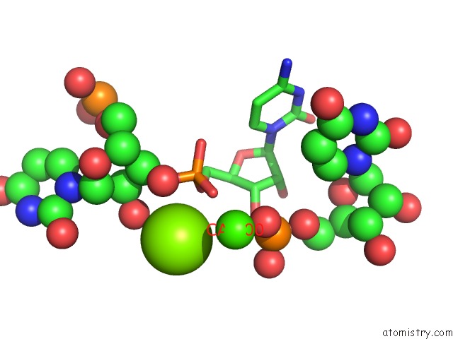
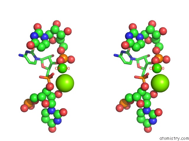
Magnesium binding site 2 out of 4 in 2hom
Go back to
Magnesium binding site 2 out
of 4 in the Crystal Structure of An E. Coli Thi-Box Riboswitch Bound to Thiamine Monophosphate
Mono view
Stereo pair view
Mono view
Stereo pair view
A full contact list of Magnesium with other atoms in the Mg binding
site number 2 of Crystal Structure of An E. Coli Thi-Box Riboswitch Bound to Thiamine Monophosphate within 5.0Å range:
|
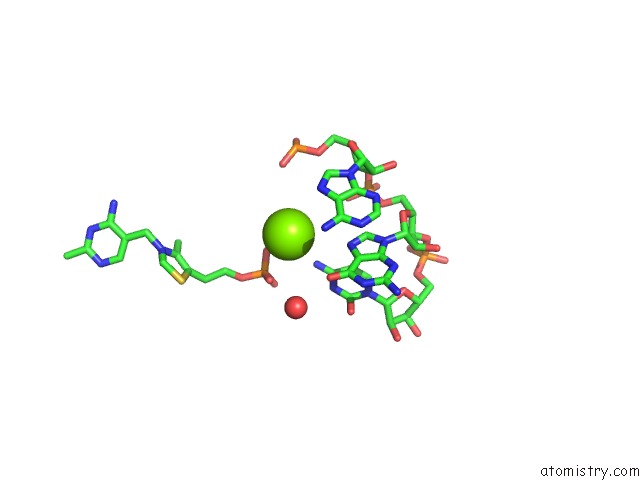
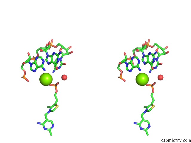
Magnesium binding site 3 out of 4 in 2hom
Go back to
Magnesium binding site 3 out
of 4 in the Crystal Structure of An E. Coli Thi-Box Riboswitch Bound to Thiamine Monophosphate
Mono view
Stereo pair view
Mono view
Stereo pair view
A full contact list of Magnesium with other atoms in the Mg binding
site number 3 of Crystal Structure of An E. Coli Thi-Box Riboswitch Bound to Thiamine Monophosphate within 5.0Å range:
|
Magnesium binding site 4 out of 4 in 2hom
Go back to
Magnesium binding site 4 out
of 4 in the Crystal Structure of An E. Coli Thi-Box Riboswitch Bound to Thiamine Monophosphate
Mono view
Stereo pair view
Mono view
Stereo pair view
A full contact list of Magnesium with other atoms in the Mg binding
site number 4 of Crystal Structure of An E. Coli Thi-Box Riboswitch Bound to Thiamine Monophosphate within 5.0Å range:
|
Reference:
T.E.Edwards,
A.R.Ferre-D'amare.
Crystal Structures of the Thi-Box Riboswitch Bound to Thiamine Pyrophosphate Analogs Reveal Adaptive Rna-Small Molecule Recognition Structure V. 14 1459 2006.
ISSN: ISSN 0969-2126
PubMed: 16962976
DOI: 10.1016/J.STR.2006.07.008
Page generated: Sun Aug 10 11:25:58 2025
ISSN: ISSN 0969-2126
PubMed: 16962976
DOI: 10.1016/J.STR.2006.07.008
Last articles
Mg in 5T4YMg in 5T63
Mg in 5T5I
Mg in 5T5C
Mg in 5T3R
Mg in 5T40
Mg in 5T41
Mg in 5T45
Mg in 5T3K
Mg in 5T14