Magnesium »
PDB 2hxh-2iea »
2i4h »
Magnesium in PDB 2i4h: Structural Studies of Protein Tyrosine Phosphatase Beta Catalytic Domain Co-Crystallized with A Sulfamic Acid Inhibitor
Enzymatic activity of Structural Studies of Protein Tyrosine Phosphatase Beta Catalytic Domain Co-Crystallized with A Sulfamic Acid Inhibitor
All present enzymatic activity of Structural Studies of Protein Tyrosine Phosphatase Beta Catalytic Domain Co-Crystallized with A Sulfamic Acid Inhibitor:
3.1.3.48;
3.1.3.48;
Protein crystallography data
The structure of Structural Studies of Protein Tyrosine Phosphatase Beta Catalytic Domain Co-Crystallized with A Sulfamic Acid Inhibitor, PDB code: 2i4h
was solved by
A.G.Evdokimov,
M.E.Pokross,
R.L.Walter,
M.Mekel,
with X-Ray Crystallography technique. A brief refinement statistics is given in the table below:
| Resolution Low / High (Å) | 27.37 / 2.15 |
| Space group | C 1 2 1 |
| Cell size a, b, c (Å), α, β, γ (°) | 113.313, 38.795, 66.983, 90.00, 104.94, 90.00 |
| R / Rfree (%) | 16.9 / 24.6 |
Other elements in 2i4h:
The structure of Structural Studies of Protein Tyrosine Phosphatase Beta Catalytic Domain Co-Crystallized with A Sulfamic Acid Inhibitor also contains other interesting chemical elements:
| Chlorine | (Cl) | 1 atom |
Magnesium Binding Sites:
The binding sites of Magnesium atom in the Structural Studies of Protein Tyrosine Phosphatase Beta Catalytic Domain Co-Crystallized with A Sulfamic Acid Inhibitor
(pdb code 2i4h). This binding sites where shown within
5.0 Angstroms radius around Magnesium atom.
In total only one binding site of Magnesium was determined in the Structural Studies of Protein Tyrosine Phosphatase Beta Catalytic Domain Co-Crystallized with A Sulfamic Acid Inhibitor, PDB code: 2i4h:
In total only one binding site of Magnesium was determined in the Structural Studies of Protein Tyrosine Phosphatase Beta Catalytic Domain Co-Crystallized with A Sulfamic Acid Inhibitor, PDB code: 2i4h:
Magnesium binding site 1 out of 1 in 2i4h
Go back to
Magnesium binding site 1 out
of 1 in the Structural Studies of Protein Tyrosine Phosphatase Beta Catalytic Domain Co-Crystallized with A Sulfamic Acid Inhibitor
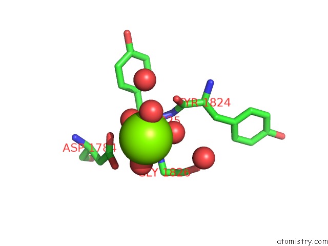
Mono view
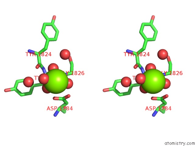
Stereo pair view
Mono view
Stereo pair view
A full contact list of Magnesium with other atoms in the Mg binding
site number 1 of Structural Studies of Protein Tyrosine Phosphatase Beta Catalytic Domain Co-Crystallized with A Sulfamic Acid Inhibitor within 5.0Å range:
|
Reference:
A.G.Evdokimov,
M.Pokross,
R.Walter,
M.Mekel,
B.Cox,
C.Li,
R.Bechard,
F.Genbauffe,
R.Andrews,
C.Diven,
B.Howard,
V.Rastogi,
J.Gray,
M.Maier,
K.G.Peters.
Engineering the Catalytic Domain of Human Protein Tyrosine Phosphatase Beta For Structure-Based Drug Discovery. Acta Crystallogr.,Sect.D V. 62 1435 2006.
ISSN: ISSN 0907-4449
PubMed: 17139078
DOI: 10.1107/S0907444906037784
Page generated: Sun Aug 10 11:31:00 2025
ISSN: ISSN 0907-4449
PubMed: 17139078
DOI: 10.1107/S0907444906037784
Last articles
Mg in 6EL4Mg in 6EL0
Mg in 6EKZ
Mg in 6EKY
Mg in 6EKX
Mg in 6EHS
Mg in 6EKW
Mg in 6EKH
Mg in 6EG5
Mg in 6EKG