Magnesium »
PDB 2hxh-2iea »
2i71 »
Magnesium in PDB 2i71: Crystal Structure of A Conserved Protein of Unknown Function From Sulfolobus Solfataricus P2
Protein crystallography data
The structure of Crystal Structure of A Conserved Protein of Unknown Function From Sulfolobus Solfataricus P2, PDB code: 2i71
was solved by
K.Tan,
T.Skarina,
O.Onopriyenko,
A.Savchenko,
A.Edwards,
A.Joachimiak,
Midwest Center For Structural Genomics (Mcsg),
with X-Ray Crystallography technique. A brief refinement statistics is given in the table below:
| Resolution Low / High (Å) | 27.92 / 1.70 |
| Space group | P 21 21 21 |
| Cell size a, b, c (Å), α, β, γ (°) | 78.849, 84.918, 108.513, 90.00, 90.00, 90.00 |
| R / Rfree (%) | 18.1 / 22.7 |
Magnesium Binding Sites:
The binding sites of Magnesium atom in the Crystal Structure of A Conserved Protein of Unknown Function From Sulfolobus Solfataricus P2
(pdb code 2i71). This binding sites where shown within
5.0 Angstroms radius around Magnesium atom.
In total 2 binding sites of Magnesium where determined in the Crystal Structure of A Conserved Protein of Unknown Function From Sulfolobus Solfataricus P2, PDB code: 2i71:
Jump to Magnesium binding site number: 1; 2;
In total 2 binding sites of Magnesium where determined in the Crystal Structure of A Conserved Protein of Unknown Function From Sulfolobus Solfataricus P2, PDB code: 2i71:
Jump to Magnesium binding site number: 1; 2;
Magnesium binding site 1 out of 2 in 2i71
Go back to
Magnesium binding site 1 out
of 2 in the Crystal Structure of A Conserved Protein of Unknown Function From Sulfolobus Solfataricus P2
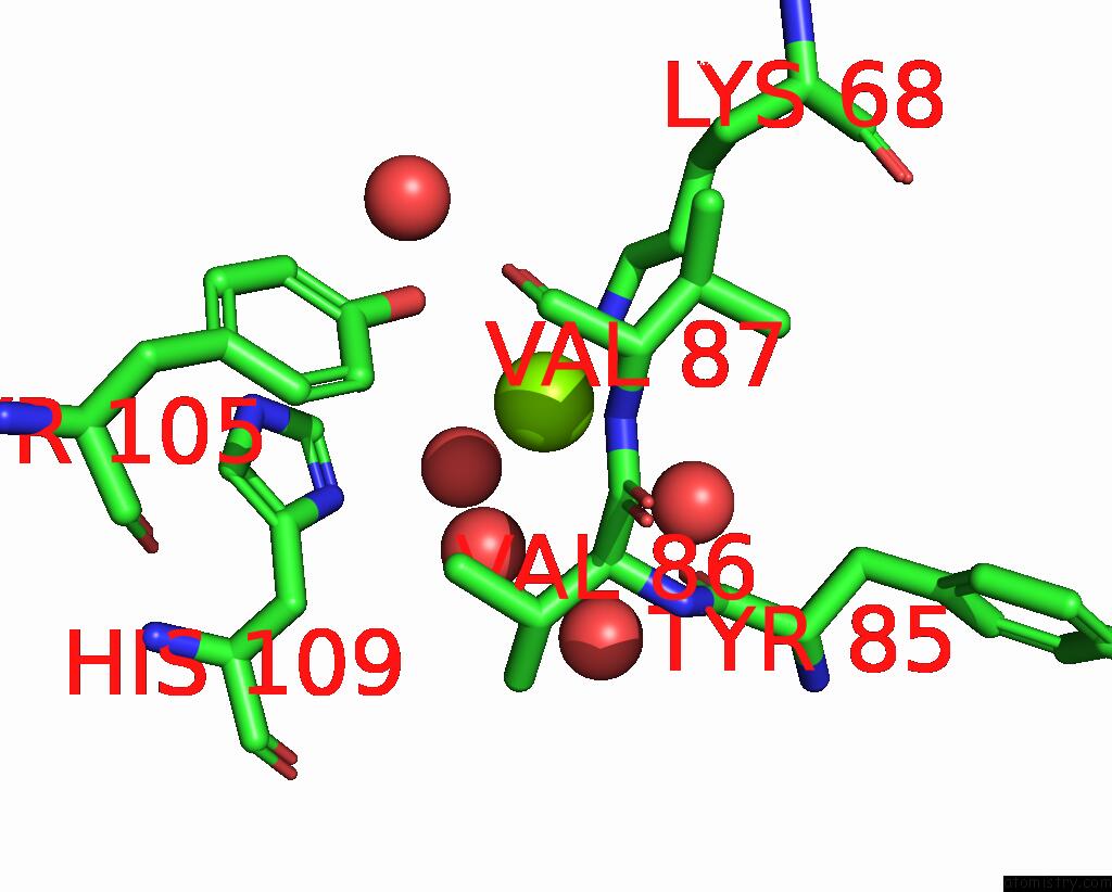
Mono view
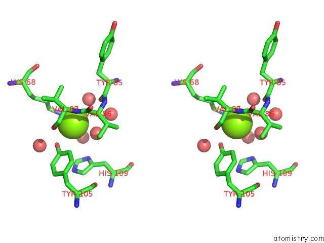
Stereo pair view
Mono view
Stereo pair view
A full contact list of Magnesium with other atoms in the Mg binding
site number 1 of Crystal Structure of A Conserved Protein of Unknown Function From Sulfolobus Solfataricus P2 within 5.0Å range:
|
Magnesium binding site 2 out of 2 in 2i71
Go back to
Magnesium binding site 2 out
of 2 in the Crystal Structure of A Conserved Protein of Unknown Function From Sulfolobus Solfataricus P2
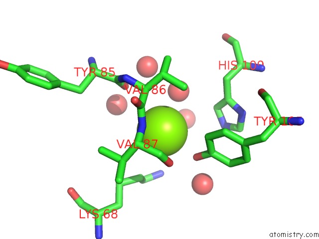
Mono view
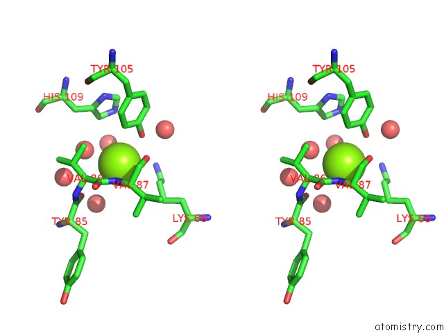
Stereo pair view
Mono view
Stereo pair view
A full contact list of Magnesium with other atoms in the Mg binding
site number 2 of Crystal Structure of A Conserved Protein of Unknown Function From Sulfolobus Solfataricus P2 within 5.0Å range:
|
Reference:
K.Tan,
T.Skarina,
O.Onopriyenko,
A.Savchenko,
A.Edwards,
A.Joachimiak.
The Crystal Structure of A Conserved Hypothetical Protein From Sulfolobus Solfataricus P2 To Be Published.
Page generated: Sun Aug 10 11:32:34 2025
Last articles
Mg in 7NASMg in 7N8M
Mg in 7N8L
Mg in 7N8E
Mg in 7N32
Mg in 7N8K
Mg in 7N6I
Mg in 7N78
Mg in 7N77
Mg in 7N73