Magnesium »
PDB 2hxh-2iea »
2iea »
Magnesium in PDB 2iea: E. Coli Pyruvate Dehydrogenase
Enzymatic activity of E. Coli Pyruvate Dehydrogenase
All present enzymatic activity of E. Coli Pyruvate Dehydrogenase:
1.2.4.1;
1.2.4.1;
Protein crystallography data
The structure of E. Coli Pyruvate Dehydrogenase, PDB code: 2iea
was solved by
W.Furey,
P.Arjunan,
with X-Ray Crystallography technique. A brief refinement statistics is given in the table below:
| Resolution Low / High (Å) | 8.00 / 1.85 |
| Space group | P 1 21 1 |
| Cell size a, b, c (Å), α, β, γ (°) | 81.690, 141.600, 82.460, 90.00, 102.40, 90.00 |
| R / Rfree (%) | 17.7 / 20.3 |
Magnesium Binding Sites:
The binding sites of Magnesium atom in the E. Coli Pyruvate Dehydrogenase
(pdb code 2iea). This binding sites where shown within
5.0 Angstroms radius around Magnesium atom.
In total 2 binding sites of Magnesium where determined in the E. Coli Pyruvate Dehydrogenase, PDB code: 2iea:
Jump to Magnesium binding site number: 1; 2;
In total 2 binding sites of Magnesium where determined in the E. Coli Pyruvate Dehydrogenase, PDB code: 2iea:
Jump to Magnesium binding site number: 1; 2;
Magnesium binding site 1 out of 2 in 2iea
Go back to
Magnesium binding site 1 out
of 2 in the E. Coli Pyruvate Dehydrogenase

Mono view
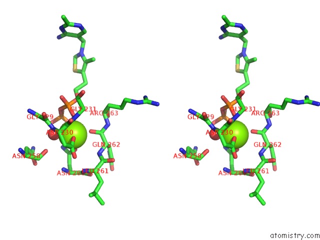
Stereo pair view
Mono view
Stereo pair view
A full contact list of Magnesium with other atoms in the Mg binding
site number 1 of E. Coli Pyruvate Dehydrogenase within 5.0Å range:
|
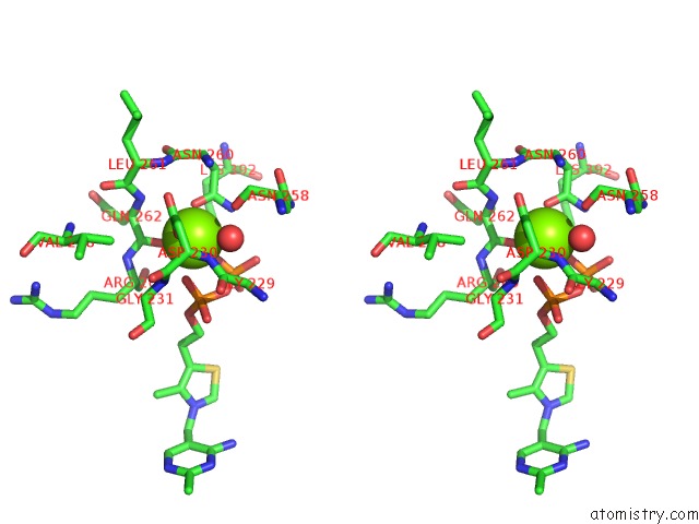
Magnesium binding site 2 out of 2 in 2iea
Go back to
Magnesium binding site 2 out
of 2 in the E. Coli Pyruvate Dehydrogenase
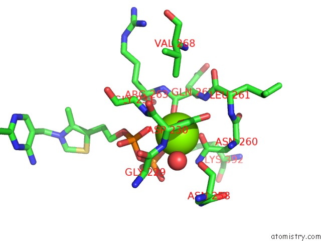
Mono view
Stereo pair view
Mono view
Stereo pair view
A full contact list of Magnesium with other atoms in the Mg binding
site number 2 of E. Coli Pyruvate Dehydrogenase within 5.0Å range:
|
Reference:
P.Arjunan,
N.Nemeria,
A.Brunskill,
K.Chandrasekhar,
M.Sax,
Y.Yan,
F.Jordan,
J.R.Guest,
W.Furey.
Structure of the Pyruvate Dehydrogenase Multienzyme Complex E1 Component From Escherichia Coli at 1.85 A Resolution. Biochemistry V. 41 5213 2002.
ISSN: ISSN 0006-2960
PubMed: 11955070
DOI: 10.1021/BI0118557
Page generated: Sun Aug 10 11:34:12 2025
ISSN: ISSN 0006-2960
PubMed: 11955070
DOI: 10.1021/BI0118557
Last articles
Mg in 2UUCMg in 2UX5
Mg in 2UX4
Mg in 2UX3
Mg in 2UWW
Mg in 2UWV
Mg in 2UWU
Mg in 2UWT
Mg in 2UUA
Mg in 2UWS