Magnesium »
PDB 2iec-2ivn »
2itn »
Magnesium in PDB 2itn: Crystal Structure of Egfr Kinase Domain G719S Mutation in Complex with Amp-Pnp
Enzymatic activity of Crystal Structure of Egfr Kinase Domain G719S Mutation in Complex with Amp-Pnp
All present enzymatic activity of Crystal Structure of Egfr Kinase Domain G719S Mutation in Complex with Amp-Pnp:
2.7.10.1;
2.7.10.1;
Protein crystallography data
The structure of Crystal Structure of Egfr Kinase Domain G719S Mutation in Complex with Amp-Pnp, PDB code: 2itn
was solved by
C.-H.Yun,
T.J.Boggon,
Y.Li,
S.Woo,
H.Greulich,
M.Meyerson,
M.J.Eck,
with X-Ray Crystallography technique. A brief refinement statistics is given in the table below:
| Resolution Low / High (Å) | 24.95 / 2.47 |
| Space group | I 2 3 |
| Cell size a, b, c (Å), α, β, γ (°) | 145.481, 145.481, 145.481, 90.00, 90.00, 90.00 |
| R / Rfree (%) | 19.7 / 26.7 |
Magnesium Binding Sites:
The binding sites of Magnesium atom in the Crystal Structure of Egfr Kinase Domain G719S Mutation in Complex with Amp-Pnp
(pdb code 2itn). This binding sites where shown within
5.0 Angstroms radius around Magnesium atom.
In total only one binding site of Magnesium was determined in the Crystal Structure of Egfr Kinase Domain G719S Mutation in Complex with Amp-Pnp, PDB code: 2itn:
In total only one binding site of Magnesium was determined in the Crystal Structure of Egfr Kinase Domain G719S Mutation in Complex with Amp-Pnp, PDB code: 2itn:
Magnesium binding site 1 out of 1 in 2itn
Go back to
Magnesium binding site 1 out
of 1 in the Crystal Structure of Egfr Kinase Domain G719S Mutation in Complex with Amp-Pnp
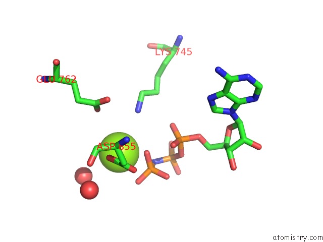
Mono view
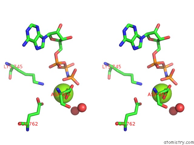
Stereo pair view
Mono view
Stereo pair view
A full contact list of Magnesium with other atoms in the Mg binding
site number 1 of Crystal Structure of Egfr Kinase Domain G719S Mutation in Complex with Amp-Pnp within 5.0Å range:
|
Reference:
C.-H.Yun,
T.J.Boggon,
Y.Li,
S.Woo,
H.Greulich,
M.Meyerson,
M.J.Eck.
Structures of Lung Cancer-Derived Egfr Mutants and Inhibitor Complexes: Mechanism of Activation and Insights Into Differential Inhibitor Sensitivity Cancer Cell V. 11 217 2007.
ISSN: ISSN 1535-6108
PubMed: 17349580
DOI: 10.1016/J.CCR.2006.12.017
Page generated: Sun Aug 10 11:40:10 2025
ISSN: ISSN 1535-6108
PubMed: 17349580
DOI: 10.1016/J.CCR.2006.12.017
Last articles
Mg in 4E7ZMg in 4E6M
Mg in 4E7S
Mg in 4E7P
Mg in 4E7O
Mg in 4E5T
Mg in 4E6N
Mg in 4E6E
Mg in 4E4P
Mg in 4E4F