Magnesium »
PDB 2iw4-2j5x »
2j0w »
Magnesium in PDB 2j0w: Crystal Structure of E. Coli Aspartokinase III in Complex with Aspartate and Adp (R-State)
Enzymatic activity of Crystal Structure of E. Coli Aspartokinase III in Complex with Aspartate and Adp (R-State)
All present enzymatic activity of Crystal Structure of E. Coli Aspartokinase III in Complex with Aspartate and Adp (R-State):
2.7.2.4;
2.7.2.4;
Protein crystallography data
The structure of Crystal Structure of E. Coli Aspartokinase III in Complex with Aspartate and Adp (R-State), PDB code: 2j0w
was solved by
M.Kotaka,
J.Ren,
M.Lockyer,
A.R.Hawkins,
D.K.Stammers,
with X-Ray Crystallography technique. A brief refinement statistics is given in the table below:
| Resolution Low / High (Å) | 29.79 / 2.5 |
| Space group | C 2 2 21 |
| Cell size a, b, c (Å), α, β, γ (°) | 49.535, 213.347, 93.078, 90.00, 90.00, 90.00 |
| R / Rfree (%) | 23.5 / 29.3 |
Other elements in 2j0w:
The structure of Crystal Structure of E. Coli Aspartokinase III in Complex with Aspartate and Adp (R-State) also contains other interesting chemical elements:
| Chlorine | (Cl) | 1 atom |
Magnesium Binding Sites:
The binding sites of Magnesium atom in the Crystal Structure of E. Coli Aspartokinase III in Complex with Aspartate and Adp (R-State)
(pdb code 2j0w). This binding sites where shown within
5.0 Angstroms radius around Magnesium atom.
In total only one binding site of Magnesium was determined in the Crystal Structure of E. Coli Aspartokinase III in Complex with Aspartate and Adp (R-State), PDB code: 2j0w:
In total only one binding site of Magnesium was determined in the Crystal Structure of E. Coli Aspartokinase III in Complex with Aspartate and Adp (R-State), PDB code: 2j0w:
Magnesium binding site 1 out of 1 in 2j0w
Go back to
Magnesium binding site 1 out
of 1 in the Crystal Structure of E. Coli Aspartokinase III in Complex with Aspartate and Adp (R-State)

Mono view
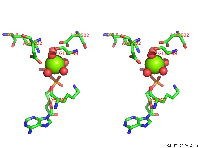
Stereo pair view
Mono view
Stereo pair view
A full contact list of Magnesium with other atoms in the Mg binding
site number 1 of Crystal Structure of E. Coli Aspartokinase III in Complex with Aspartate and Adp (R-State) within 5.0Å range:
|
Reference:
M.Kotaka,
J.Ren,
M.Lockyer,
A.R.Hawkins,
D.K.Stammers.
Structures of R- and T-State Escherichia Coli Aspartokinase III: Mechanisms of the Allosteric Transition and Inhibition By Lysine. J.Biol.Chem. V. 281 31544 2006.
ISSN: ISSN 0021-9258
PubMed: 16905770
DOI: 10.1074/JBC.M605886200
Page generated: Wed Aug 14 00:21:58 2024
ISSN: ISSN 0021-9258
PubMed: 16905770
DOI: 10.1074/JBC.M605886200
Last articles
Zn in 9J0NZn in 9J0O
Zn in 9J0P
Zn in 9FJX
Zn in 9EKB
Zn in 9C0F
Zn in 9CAH
Zn in 9CH0
Zn in 9CH3
Zn in 9CH1