Magnesium »
PDB 2iw4-2j5x »
2j17 »
Magnesium in PDB 2j17: Ptyr Bound Form of Sdp-1
Enzymatic activity of Ptyr Bound Form of Sdp-1
All present enzymatic activity of Ptyr Bound Form of Sdp-1:
3.1.3.48;
3.1.3.48;
Protein crystallography data
The structure of Ptyr Bound Form of Sdp-1, PDB code: 2j17
was solved by
D.C.Briggs,
N.Q.Mcdonald,
with X-Ray Crystallography technique. A brief refinement statistics is given in the table below:
| Resolution Low / High (Å) | 59.34 / 2.84 |
| Space group | P 43 21 2 |
| Cell size a, b, c (Å), α, β, γ (°) | 61.661, 61.661, 217.562, 90.00, 90.00, 90.00 |
| R / Rfree (%) | 22.1 / 26.9 |
Magnesium Binding Sites:
The binding sites of Magnesium atom in the Ptyr Bound Form of Sdp-1
(pdb code 2j17). This binding sites where shown within
5.0 Angstroms radius around Magnesium atom.
In total 5 binding sites of Magnesium where determined in the Ptyr Bound Form of Sdp-1, PDB code: 2j17:
Jump to Magnesium binding site number: 1; 2; 3; 4; 5;
In total 5 binding sites of Magnesium where determined in the Ptyr Bound Form of Sdp-1, PDB code: 2j17:
Jump to Magnesium binding site number: 1; 2; 3; 4; 5;
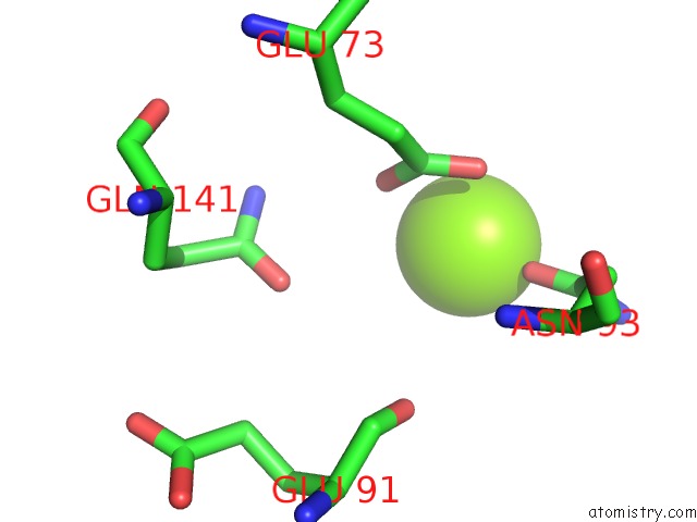
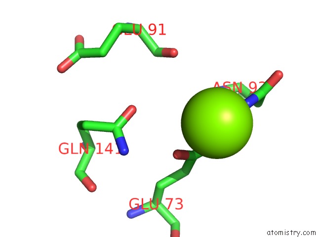
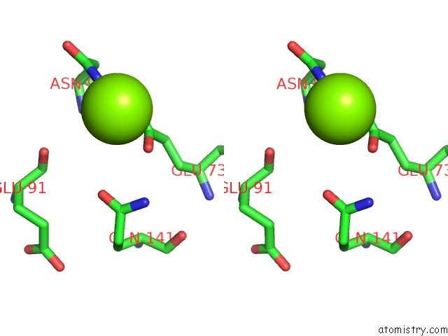
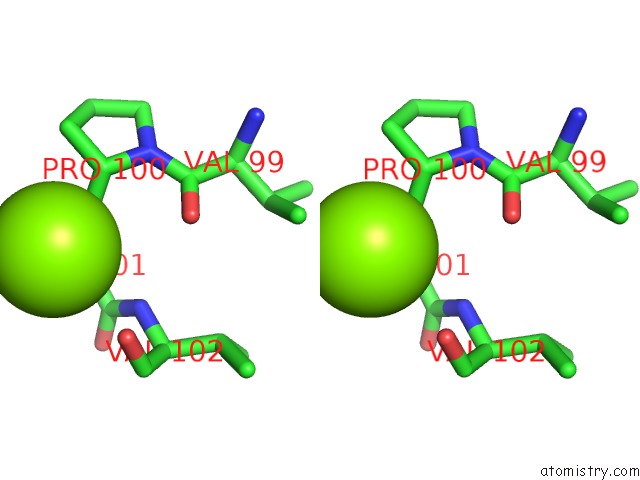
Magnesium binding site 1 out of 5 in 2j17
Go back to
Magnesium binding site 1 out
of 5 in the Ptyr Bound Form of Sdp-1
Mono view
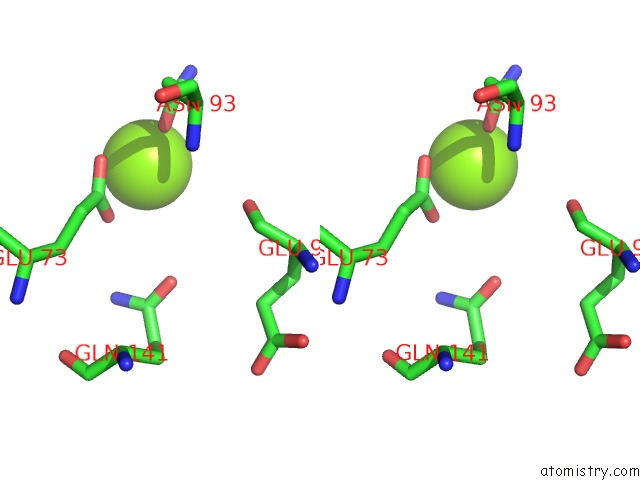
Stereo pair view
Mono view
Stereo pair view
A full contact list of Magnesium with other atoms in the Mg binding
site number 1 of Ptyr Bound Form of Sdp-1 within 5.0Å range:
|
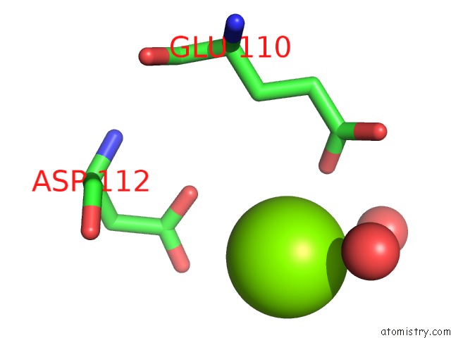
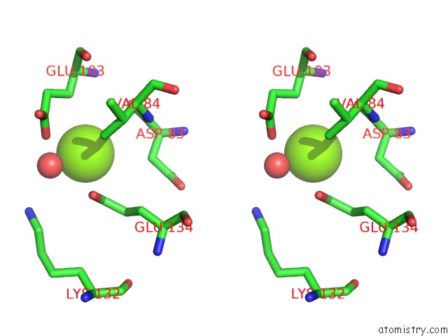
Magnesium binding site 2 out of 5 in 2j17
Go back to
Magnesium binding site 2 out
of 5 in the Ptyr Bound Form of Sdp-1
Mono view
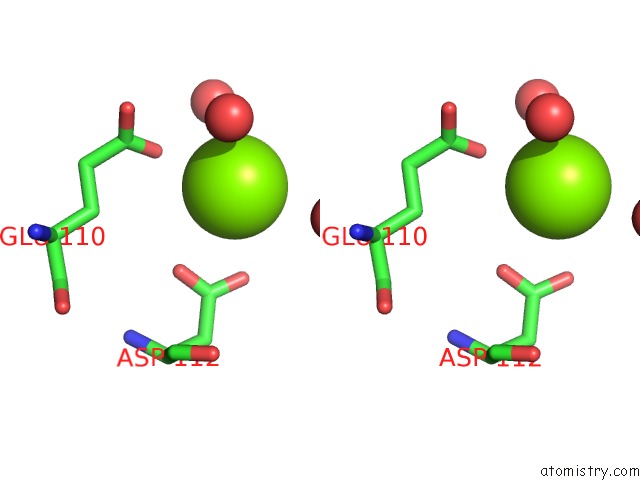
Stereo pair view
Mono view
Stereo pair view
A full contact list of Magnesium with other atoms in the Mg binding
site number 2 of Ptyr Bound Form of Sdp-1 within 5.0Å range:
|
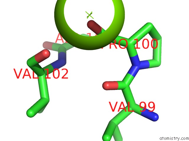
Magnesium binding site 3 out of 5 in 2j17
Go back to
Magnesium binding site 3 out
of 5 in the Ptyr Bound Form of Sdp-1
Mono view
Stereo pair view
Mono view
Stereo pair view
A full contact list of Magnesium with other atoms in the Mg binding
site number 3 of Ptyr Bound Form of Sdp-1 within 5.0Å range:
|
Magnesium binding site 4 out of 5 in 2j17
Go back to
Magnesium binding site 4 out
of 5 in the Ptyr Bound Form of Sdp-1

Mono view
Stereo pair view
Mono view
Stereo pair view
A full contact list of Magnesium with other atoms in the Mg binding
site number 4 of Ptyr Bound Form of Sdp-1 within 5.0Å range:
|
Magnesium binding site 5 out of 5 in 2j17
Go back to
Magnesium binding site 5 out
of 5 in the Ptyr Bound Form of Sdp-1
Mono view
Stereo pair view
Mono view
Stereo pair view
A full contact list of Magnesium with other atoms in the Mg binding
site number 5 of Ptyr Bound Form of Sdp-1 within 5.0Å range:
|
Reference:
G.C.Fox,
M.Shafiq,
D.C.Briggs,
P.P.Knowles,
M.Collister,
M.J.Didmon,
V.Makrantoni,
R.J.Dickinson,
S.Hanrahan,
N.Totty,
M.J.Stark,
S.M.Keyse,
N.Q.Mcdonald.
Redox-Mediated Substrate Recognition By SDP1 Defines A New Group of Tyrosine Phosphatases. Nature V. 447 487 2007.
ISSN: ESSN 1476-4687
PubMed: 17495930
DOI: 10.1038/NATURE05804
Page generated: Sun Aug 10 11:44:05 2025
ISSN: ESSN 1476-4687
PubMed: 17495930
DOI: 10.1038/NATURE05804
Last articles
Mg in 2UUCMg in 2UX5
Mg in 2UX4
Mg in 2UX3
Mg in 2UWW
Mg in 2UWV
Mg in 2UWU
Mg in 2UWT
Mg in 2UUA
Mg in 2UWS