Magnesium »
PDB 2j7k-2jg2 »
2jao »
Magnesium in PDB 2jao: Crystal Structure of D12N Variant of Mouse Cytosolic 5'(3')- Deoxyribonucleotidase (Cdn) in Complex with Deoxyguanosine 5'- Monophosphate
Protein crystallography data
The structure of Crystal Structure of D12N Variant of Mouse Cytosolic 5'(3')- Deoxyribonucleotidase (Cdn) in Complex with Deoxyguanosine 5'- Monophosphate, PDB code: 2jao
was solved by
K.Wallden,
B.Ruzzenente,
V.Bianchi,
P.Nordlund,
with X-Ray Crystallography technique. A brief refinement statistics is given in the table below:
| Resolution Low / High (Å) | 66.08 / 2.00 |
| Space group | P 62 |
| Cell size a, b, c (Å), α, β, γ (°) | 76.250, 76.250, 85.760, 90.00, 90.00, 120.00 |
| R / Rfree (%) | 16.5 / 20.5 |
Magnesium Binding Sites:
The binding sites of Magnesium atom in the Crystal Structure of D12N Variant of Mouse Cytosolic 5'(3')- Deoxyribonucleotidase (Cdn) in Complex with Deoxyguanosine 5'- Monophosphate
(pdb code 2jao). This binding sites where shown within
5.0 Angstroms radius around Magnesium atom.
In total only one binding site of Magnesium was determined in the Crystal Structure of D12N Variant of Mouse Cytosolic 5'(3')- Deoxyribonucleotidase (Cdn) in Complex with Deoxyguanosine 5'- Monophosphate, PDB code: 2jao:
In total only one binding site of Magnesium was determined in the Crystal Structure of D12N Variant of Mouse Cytosolic 5'(3')- Deoxyribonucleotidase (Cdn) in Complex with Deoxyguanosine 5'- Monophosphate, PDB code: 2jao:
Magnesium binding site 1 out of 1 in 2jao
Go back to
Magnesium binding site 1 out
of 1 in the Crystal Structure of D12N Variant of Mouse Cytosolic 5'(3')- Deoxyribonucleotidase (Cdn) in Complex with Deoxyguanosine 5'- Monophosphate
Mono view
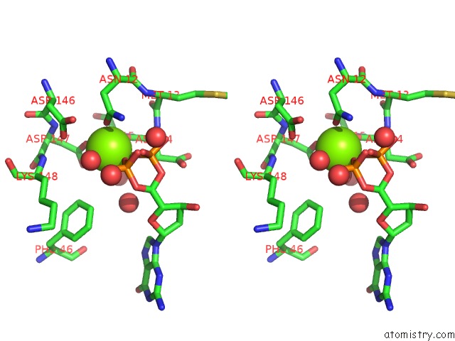
Stereo pair view
Mono view
Stereo pair view
A full contact list of Magnesium with other atoms in the Mg binding
site number 1 of Crystal Structure of D12N Variant of Mouse Cytosolic 5'(3')- Deoxyribonucleotidase (Cdn) in Complex with Deoxyguanosine 5'- Monophosphate within 5.0Å range:
|
Reference:
K.Wallden,
A.Rinaldo-Matthis,
B.Ruzzenente,
C.Rampazzo,
V.Bianchi,
P.Nordlund.
Crystal Structures of Human and Murine Deoxyribonucleotidases: Insights Into Recognition of Substrates and Nucleotide Analogues. Biochemistry V. 46 13809 2007.
ISSN: ISSN 0006-2960
PubMed: 17985935
DOI: 10.1021/BI7014794
Page generated: Wed Aug 14 00:41:32 2024
ISSN: ISSN 0006-2960
PubMed: 17985935
DOI: 10.1021/BI7014794
Last articles
Fe in 2YXOFe in 2YRS
Fe in 2YXC
Fe in 2YNM
Fe in 2YVJ
Fe in 2YP1
Fe in 2YU2
Fe in 2YU1
Fe in 2YQB
Fe in 2YOO