Magnesium »
PDB 2j7k-2jg2 »
2jau »
Magnesium in PDB 2jau: Crystal Structure of D41N Variant of Human Mitochondrial 5'(3')- Deoxyribonucleotidase (Mdn) in Complex with 3'-Azidothymidine 5'- Monophosphate
Protein crystallography data
The structure of Crystal Structure of D41N Variant of Human Mitochondrial 5'(3')- Deoxyribonucleotidase (Mdn) in Complex with 3'-Azidothymidine 5'- Monophosphate, PDB code: 2jau
was solved by
K.Wallden,
B.Ruzzenente,
V.Bianchi,
P.Nordlund,
with X-Ray Crystallography technique. A brief refinement statistics is given in the table below:
| Resolution Low / High (Å) | 40.00 / 1.80 |
| Space group | P 43 21 2 |
| Cell size a, b, c (Å), α, β, γ (°) | 73.590, 73.590, 105.740, 90.00, 90.00, 90.00 |
| R / Rfree (%) | 18.2 / 21.4 |
Magnesium Binding Sites:
The binding sites of Magnesium atom in the Crystal Structure of D41N Variant of Human Mitochondrial 5'(3')- Deoxyribonucleotidase (Mdn) in Complex with 3'-Azidothymidine 5'- Monophosphate
(pdb code 2jau). This binding sites where shown within
5.0 Angstroms radius around Magnesium atom.
In total 2 binding sites of Magnesium where determined in the Crystal Structure of D41N Variant of Human Mitochondrial 5'(3')- Deoxyribonucleotidase (Mdn) in Complex with 3'-Azidothymidine 5'- Monophosphate, PDB code: 2jau:
Jump to Magnesium binding site number: 1; 2;
In total 2 binding sites of Magnesium where determined in the Crystal Structure of D41N Variant of Human Mitochondrial 5'(3')- Deoxyribonucleotidase (Mdn) in Complex with 3'-Azidothymidine 5'- Monophosphate, PDB code: 2jau:
Jump to Magnesium binding site number: 1; 2;
Magnesium binding site 1 out of 2 in 2jau
Go back to
Magnesium binding site 1 out
of 2 in the Crystal Structure of D41N Variant of Human Mitochondrial 5'(3')- Deoxyribonucleotidase (Mdn) in Complex with 3'-Azidothymidine 5'- Monophosphate
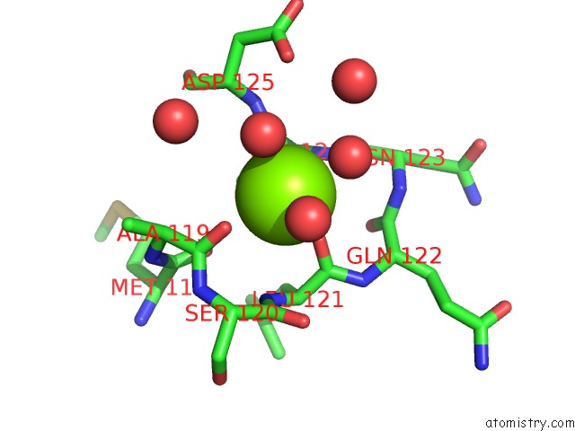
Mono view
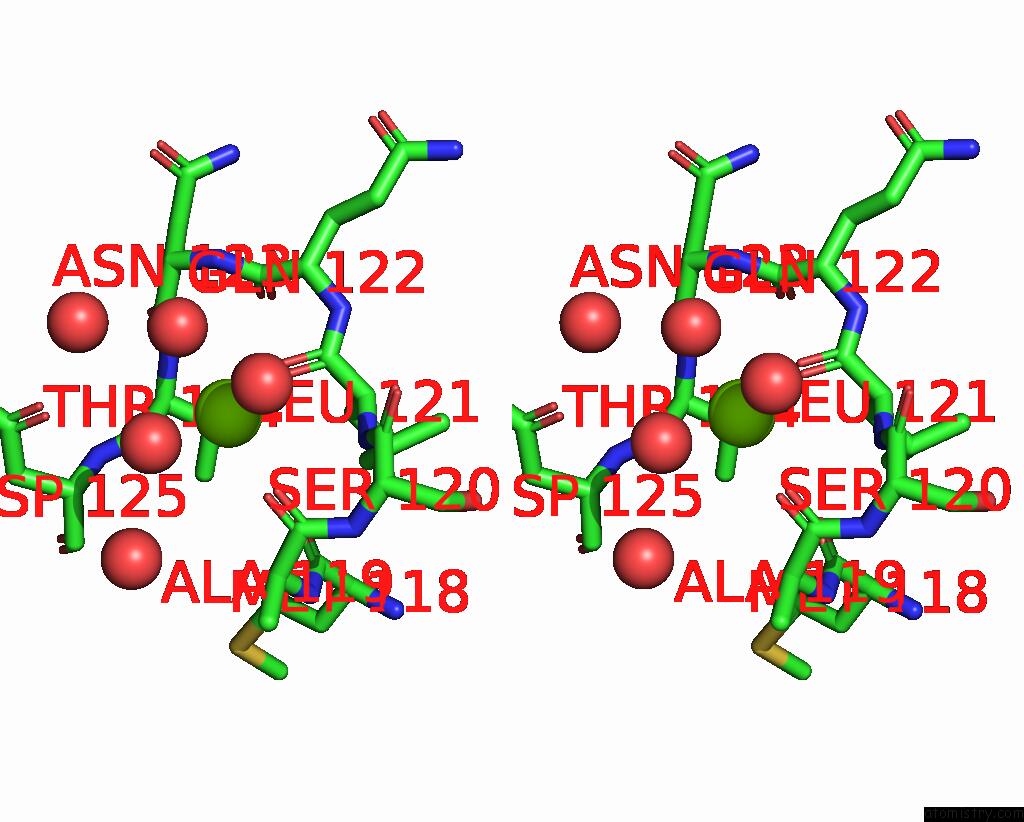
Stereo pair view
Mono view
Stereo pair view
A full contact list of Magnesium with other atoms in the Mg binding
site number 1 of Crystal Structure of D41N Variant of Human Mitochondrial 5'(3')- Deoxyribonucleotidase (Mdn) in Complex with 3'-Azidothymidine 5'- Monophosphate within 5.0Å range:
|
Magnesium binding site 2 out of 2 in 2jau
Go back to
Magnesium binding site 2 out
of 2 in the Crystal Structure of D41N Variant of Human Mitochondrial 5'(3')- Deoxyribonucleotidase (Mdn) in Complex with 3'-Azidothymidine 5'- Monophosphate
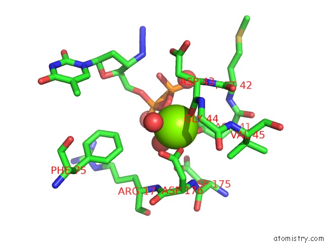
Mono view
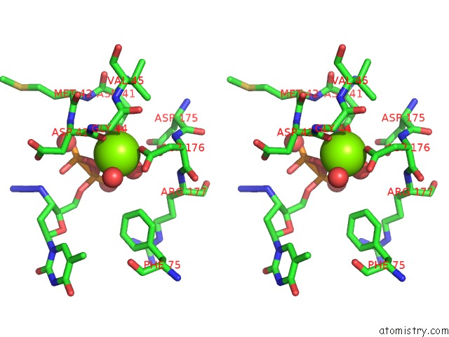
Stereo pair view
Mono view
Stereo pair view
A full contact list of Magnesium with other atoms in the Mg binding
site number 2 of Crystal Structure of D41N Variant of Human Mitochondrial 5'(3')- Deoxyribonucleotidase (Mdn) in Complex with 3'-Azidothymidine 5'- Monophosphate within 5.0Å range:
|
Reference:
K.Wallden,
A.Rinaldo-Matthis,
B.Ruzzenente,
C.Rampazzo,
V.Bianchi,
P.Nordlund.
Crystal Structures of Human and Murine Deoxyribonucleotidases: Insights Into Recognition of Substrates and Nucleotide Analogues. Biochemistry V. 46 13809 2007.
ISSN: ISSN 0006-2960
PubMed: 17985935
DOI: 10.1021/BI7014794
Page generated: Sun Aug 10 11:56:27 2025
ISSN: ISSN 0006-2960
PubMed: 17985935
DOI: 10.1021/BI7014794
Last articles
Mg in 5G0RMg in 5G5V
Mg in 5G3T
Mg in 5G5T
Mg in 5G5S
Mg in 5G4A
Mg in 5G57
Mg in 5G50
Mg in 5G41
Mg in 5G3Z