Magnesium »
PDB 2j7k-2jg2 »
2jc9 »
Magnesium in PDB 2jc9: Crystal Structure of Human Cytosolic 5'-Nucleotidase II in Complex with Adenosine
Enzymatic activity of Crystal Structure of Human Cytosolic 5'-Nucleotidase II in Complex with Adenosine
All present enzymatic activity of Crystal Structure of Human Cytosolic 5'-Nucleotidase II in Complex with Adenosine:
3.1.3.5;
3.1.3.5;
Protein crystallography data
The structure of Crystal Structure of Human Cytosolic 5'-Nucleotidase II in Complex with Adenosine, PDB code: 2jc9
was solved by
K.Wallden,
P.Stenmark,
C.Arrowsmith,
H.Berglund,
R.Busam,
R.Collins,
A.Edwards,
M.Ehn,
S.Flodin,
A.Flores,
S.Graslund,
M.Hammarstrom,
B.M.Hallberg,
S.L.Holmberg,
M.Hogbom,
T.Karlberg,
T.Kotenyova,
A.Magnusdottir,
P.Nilsson-Ehle,
T.Nyman,
D.Ogg,
C.Persson,
J.Sagemark,
M.Sundstrom,
J.Uppenberg,
A.G.Thorsell,
S.Van Den Berg,
P.Loppnau,
J.Weigelt,
M.Welin,
P.Nordlund,
with X-Ray Crystallography technique. A brief refinement statistics is given in the table below:
| Resolution Low / High (Å) | 74.33 / 1.5 |
| Space group | I 2 2 2 |
| Cell size a, b, c (Å), α, β, γ (°) | 91.273, 127.983, 130.601, 90.00, 90.00, 90.00 |
| R / Rfree (%) | 16 / 18.6 |
Magnesium Binding Sites:
The binding sites of Magnesium atom in the Crystal Structure of Human Cytosolic 5'-Nucleotidase II in Complex with Adenosine
(pdb code 2jc9). This binding sites where shown within
5.0 Angstroms radius around Magnesium atom.
In total only one binding site of Magnesium was determined in the Crystal Structure of Human Cytosolic 5'-Nucleotidase II in Complex with Adenosine, PDB code: 2jc9:
In total only one binding site of Magnesium was determined in the Crystal Structure of Human Cytosolic 5'-Nucleotidase II in Complex with Adenosine, PDB code: 2jc9:
Magnesium binding site 1 out of 1 in 2jc9
Go back to
Magnesium binding site 1 out
of 1 in the Crystal Structure of Human Cytosolic 5'-Nucleotidase II in Complex with Adenosine
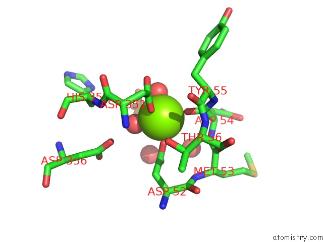
Mono view
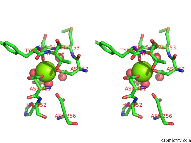
Stereo pair view
Mono view
Stereo pair view
A full contact list of Magnesium with other atoms in the Mg binding
site number 1 of Crystal Structure of Human Cytosolic 5'-Nucleotidase II in Complex with Adenosine within 5.0Å range:
|
Reference:
K.Wallden,
P.Stenmark,
T.Nyman,
S.Flodin,
S.Graslund,
P.Loppnau,
V.Bianchi,
P.Nordlund.
Crystal Structure of Human Cytosolic 5'- Nucleotidase II: Insights Into Allosteric Regulation and Substrate Recognition J.Biol.Chem. V. 282 17828 2007.
ISSN: ISSN 0021-9258
PubMed: 17405878
DOI: 10.1074/JBC.M700917200
Page generated: Sun Aug 10 11:57:15 2025
ISSN: ISSN 0021-9258
PubMed: 17405878
DOI: 10.1074/JBC.M700917200
Last articles
Mg in 2VSCMg in 2VPN
Mg in 2VRN
Mg in 2VPQ
Mg in 2VQD
Mg in 2VQ2
Mg in 2VPR
Mg in 2VPO
Mg in 2VP0
Mg in 2VOS