Magnesium »
PDB 2jga-2mse »
2jga »
Magnesium in PDB 2jga: Crystal Structure of Human Cytosolic 5'-Nucleotidase III in Complex with Phosphate and Magnesium
Enzymatic activity of Crystal Structure of Human Cytosolic 5'-Nucleotidase III in Complex with Phosphate and Magnesium
All present enzymatic activity of Crystal Structure of Human Cytosolic 5'-Nucleotidase III in Complex with Phosphate and Magnesium:
3.1.3.5;
3.1.3.5;
Protein crystallography data
The structure of Crystal Structure of Human Cytosolic 5'-Nucleotidase III in Complex with Phosphate and Magnesium, PDB code: 2jga
was solved by
K.Wallden,
P.Stenmark,
C.Arrowsmith,
H.Berglund,
R.Collins,
A.Edwards,
M.Ehn,
S.Flodin,
A.Flores,
S.Graslund,
M.Hammarstrom,
M.Hallberg,
B.Holmberg,
L.Schiavone,
M.Hogbom,
T.Kotenyova,
A.Magnusdottir,
P.Nilsson-Ehle,
T.Nyman,
D.Ogg,
C.Persson,
J.Sagemark,
M.Sundstrom,
A.G.Thorsell,
J.Uppenberg,
S.Van Den Berg,
J.Weigelt,
M.Welin,
P.Nordlund,
with X-Ray Crystallography technique. A brief refinement statistics is given in the table below:
| Resolution Low / High (Å) | 50.19 / 3.01 |
| Space group | C 2 2 21 |
| Cell size a, b, c (Å), α, β, γ (°) | 87.459, 101.036, 77.046, 90.00, 90.00, 90.00 |
| R / Rfree (%) | 18.1 / 26.8 |
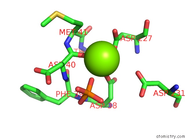
Magnesium Binding Sites:
The binding sites of Magnesium atom in the Crystal Structure of Human Cytosolic 5'-Nucleotidase III in Complex with Phosphate and Magnesium
(pdb code 2jga). This binding sites where shown within
5.0 Angstroms radius around Magnesium atom.
In total only one binding site of Magnesium was determined in the Crystal Structure of Human Cytosolic 5'-Nucleotidase III in Complex with Phosphate and Magnesium, PDB code: 2jga:
In total only one binding site of Magnesium was determined in the Crystal Structure of Human Cytosolic 5'-Nucleotidase III in Complex with Phosphate and Magnesium, PDB code: 2jga:
Magnesium binding site 1 out of 1 in 2jga
Go back to
Magnesium binding site 1 out
of 1 in the Crystal Structure of Human Cytosolic 5'-Nucleotidase III in Complex with Phosphate and Magnesium
Mono view
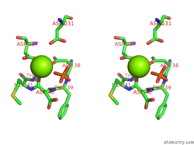
Stereo pair view
Mono view
Stereo pair view
A full contact list of Magnesium with other atoms in the Mg binding
site number 1 of Crystal Structure of Human Cytosolic 5'-Nucleotidase III in Complex with Phosphate and Magnesium within 5.0Å range:
|
Reference:
K.Wallden,
P.Stenmark,
T.Nyman,
S.Flodin,
S.Graslund,
P.Loppnau,
V.Bianchi,
P.Nordlund.
Crystal Structure of Human Cytosolic 5'-Nucleotidase II: Insights Into Allosteric Regulation and Substrate Recognition. J.Biol.Chem. V. 282 17828 2007.
ISSN: ISSN 0021-9258
PubMed: 17405878
DOI: 10.1074/JBC.M700917200
Page generated: Wed Aug 14 00:45:56 2024
ISSN: ISSN 0021-9258
PubMed: 17405878
DOI: 10.1074/JBC.M700917200
Last articles
Zn in 9MJ5Zn in 9HNW
Zn in 9G0L
Zn in 9FNE
Zn in 9DZN
Zn in 9E0I
Zn in 9D32
Zn in 9DAK
Zn in 8ZXC
Zn in 8ZUF