Magnesium »
PDB 2jga-2mse »
2jhr »
Magnesium in PDB 2jhr: Crystal Structure of Myosin-2 Motor Domain in Complex with Adp- Metavanadate and Pentabromopseudilin
Protein crystallography data
The structure of Crystal Structure of Myosin-2 Motor Domain in Complex with Adp- Metavanadate and Pentabromopseudilin, PDB code: 2jhr
was solved by
R.Fedorov,
M.Boehl,
G.Tsiavaliaris,
F.K.Hartmann,
P.Baruch,
B.Brenner,
R.Martin,
H.J.Knoelker,
H.O.Gutzeit,
D.J.Manstein,
with X-Ray Crystallography technique. A brief refinement statistics is given in the table below:
| Resolution Low / High (Å) | 8.00 / 2.80 |
| Space group | C 2 2 21 |
| Cell size a, b, c (Å), α, β, γ (°) | 89.758, 150.464, 154.550, 90.00, 90.00, 90.00 |
| R / Rfree (%) | 21.7 / 26.5 |
Other elements in 2jhr:
The structure of Crystal Structure of Myosin-2 Motor Domain in Complex with Adp- Metavanadate and Pentabromopseudilin also contains other interesting chemical elements:
| Bromine | (Br) | 5 atoms |
| Vanadium | (V) | 1 atom |
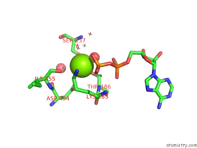
Magnesium Binding Sites:
The binding sites of Magnesium atom in the Crystal Structure of Myosin-2 Motor Domain in Complex with Adp- Metavanadate and Pentabromopseudilin
(pdb code 2jhr). This binding sites where shown within
5.0 Angstroms radius around Magnesium atom.
In total only one binding site of Magnesium was determined in the Crystal Structure of Myosin-2 Motor Domain in Complex with Adp- Metavanadate and Pentabromopseudilin, PDB code: 2jhr:
In total only one binding site of Magnesium was determined in the Crystal Structure of Myosin-2 Motor Domain in Complex with Adp- Metavanadate and Pentabromopseudilin, PDB code: 2jhr:
Magnesium binding site 1 out of 1 in 2jhr
Go back to
Magnesium binding site 1 out
of 1 in the Crystal Structure of Myosin-2 Motor Domain in Complex with Adp- Metavanadate and Pentabromopseudilin
Mono view

Stereo pair view
Mono view
Stereo pair view
A full contact list of Magnesium with other atoms in the Mg binding
site number 1 of Crystal Structure of Myosin-2 Motor Domain in Complex with Adp- Metavanadate and Pentabromopseudilin within 5.0Å range:
|
Reference:
R.Fedorov,
M.Bohl,
G.Tsiavaliaris,
F.K.Hartmann,
M.H.Taft,
P.Baruch,
B.Brenner,
R.Martin,
H.Knolker,
H.O.Gutzeit,
D.J.Manstein.
The Mechanism of Pentabromopseudilin Inhibition of Myosin Motor Activity. Nat.Struct.Mol.Biol. V. 16 80 2009.
ISSN: ISSN 1545-9993
PubMed: 19122661
DOI: 10.1038/NSMB.1542
Page generated: Sun Aug 10 11:59:11 2025
ISSN: ISSN 1545-9993
PubMed: 19122661
DOI: 10.1038/NSMB.1542
Last articles
Mg in 3C4JMg in 3C41
Mg in 3C3I
Mg in 3C1M
Mg in 3C3C
Mg in 3C2T
Mg in 3C3A
Mg in 3C15
Mg in 3C16
Mg in 3C14