Magnesium »
PDB 2jga-2mse »
2ji9 »
Magnesium in PDB 2ji9: X-Ray Structure of Oxalyl-Coa Decarboxylase in Complex with 3-Deaza-Thdp
Enzymatic activity of X-Ray Structure of Oxalyl-Coa Decarboxylase in Complex with 3-Deaza-Thdp
All present enzymatic activity of X-Ray Structure of Oxalyl-Coa Decarboxylase in Complex with 3-Deaza-Thdp:
4.1.1.8;
4.1.1.8;
Protein crystallography data
The structure of X-Ray Structure of Oxalyl-Coa Decarboxylase in Complex with 3-Deaza-Thdp, PDB code: 2ji9
was solved by
C.L.Berthold,
C.G.Toyota,
P.Moussatche,
M.D.Wood,
F.Leeper,
N.G.J.Richards,
Y.Lindqvist,
with X-Ray Crystallography technique. A brief refinement statistics is given in the table below:
| Resolution Low / High (Å) | 30.00 / 2.2 |
| Space group | P 31 2 1 |
| Cell size a, b, c (Å), α, β, γ (°) | 127.721, 127.721, 152.417, 90.00, 90.00, 120.00 |
| R / Rfree (%) | 17.7 / 21.5 |
Magnesium Binding Sites:
The binding sites of Magnesium atom in the X-Ray Structure of Oxalyl-Coa Decarboxylase in Complex with 3-Deaza-Thdp
(pdb code 2ji9). This binding sites where shown within
5.0 Angstroms radius around Magnesium atom.
In total 2 binding sites of Magnesium where determined in the X-Ray Structure of Oxalyl-Coa Decarboxylase in Complex with 3-Deaza-Thdp, PDB code: 2ji9:
Jump to Magnesium binding site number: 1; 2;
In total 2 binding sites of Magnesium where determined in the X-Ray Structure of Oxalyl-Coa Decarboxylase in Complex with 3-Deaza-Thdp, PDB code: 2ji9:
Jump to Magnesium binding site number: 1; 2;
Magnesium binding site 1 out of 2 in 2ji9
Go back to
Magnesium binding site 1 out
of 2 in the X-Ray Structure of Oxalyl-Coa Decarboxylase in Complex with 3-Deaza-Thdp

Mono view
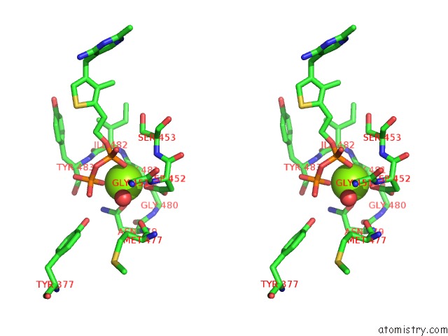
Stereo pair view
Mono view
Stereo pair view
A full contact list of Magnesium with other atoms in the Mg binding
site number 1 of X-Ray Structure of Oxalyl-Coa Decarboxylase in Complex with 3-Deaza-Thdp within 5.0Å range:
|
Magnesium binding site 2 out of 2 in 2ji9
Go back to
Magnesium binding site 2 out
of 2 in the X-Ray Structure of Oxalyl-Coa Decarboxylase in Complex with 3-Deaza-Thdp

Mono view

Stereo pair view
Mono view
Stereo pair view
A full contact list of Magnesium with other atoms in the Mg binding
site number 2 of X-Ray Structure of Oxalyl-Coa Decarboxylase in Complex with 3-Deaza-Thdp within 5.0Å range:
|
Reference:
C.L.Berthold,
C.G.Toyota,
P.Moussatche,
M.D.Wood,
F.Leeper,
N.G.J.Richards,
Y.Lindqvist.
Crystallographic Snapshots of Oxalyl-Coa Decarboxylase Give Insights Into Catalysis By Nonoxidative Thdp-Dependent Decarboxylases Structure V. 15 853 2007.
ISSN: ISSN 0969-2126
PubMed: 17637344
DOI: 10.1016/J.STR.2007.06.001
Page generated: Wed Aug 14 00:45:57 2024
ISSN: ISSN 0969-2126
PubMed: 17637344
DOI: 10.1016/J.STR.2007.06.001
Last articles
Zn in 9MJ5Zn in 9HNW
Zn in 9G0L
Zn in 9FNE
Zn in 9DZN
Zn in 9E0I
Zn in 9D32
Zn in 9DAK
Zn in 8ZXC
Zn in 8ZUF