Magnesium »
PDB 2jga-2mse »
2jj9 »
Magnesium in PDB 2jj9: Crystal Structure of Myosin-2 in Complex with Adp-Metavanadate
Protein crystallography data
The structure of Crystal Structure of Myosin-2 in Complex with Adp-Metavanadate, PDB code: 2jj9
was solved by
R.Fedorov,
M.Boehl,
G.Tsiavaliaris,
F.K.Hartmann,
P.Baruch,
B.Brenner,
R.Martin,
H.J.Knoelker,
H.O.Gutzeit,
D.J.Manstein,
with X-Ray Crystallography technique. A brief refinement statistics is given in the table below:
| Resolution Low / High (Å) | 8.00 / 2.30 |
| Space group | C 2 2 21 |
| Cell size a, b, c (Å), α, β, γ (°) | 89.417, 147.218, 153.837, 90.00, 90.00, 90.00 |
| R / Rfree (%) | 22.5 / 27.5 |
Other elements in 2jj9:
The structure of Crystal Structure of Myosin-2 in Complex with Adp-Metavanadate also contains other interesting chemical elements:
| Vanadium | (V) | 1 atom |
Magnesium Binding Sites:
The binding sites of Magnesium atom in the Crystal Structure of Myosin-2 in Complex with Adp-Metavanadate
(pdb code 2jj9). This binding sites where shown within
5.0 Angstroms radius around Magnesium atom.
In total only one binding site of Magnesium was determined in the Crystal Structure of Myosin-2 in Complex with Adp-Metavanadate, PDB code: 2jj9:
In total only one binding site of Magnesium was determined in the Crystal Structure of Myosin-2 in Complex with Adp-Metavanadate, PDB code: 2jj9:
Magnesium binding site 1 out of 1 in 2jj9
Go back to
Magnesium binding site 1 out
of 1 in the Crystal Structure of Myosin-2 in Complex with Adp-Metavanadate
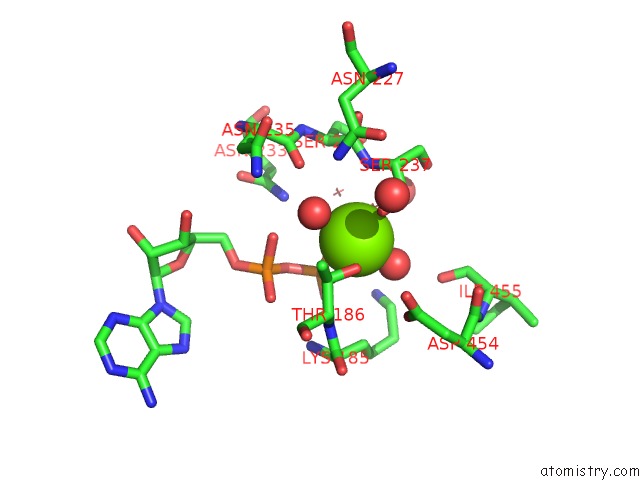
Mono view
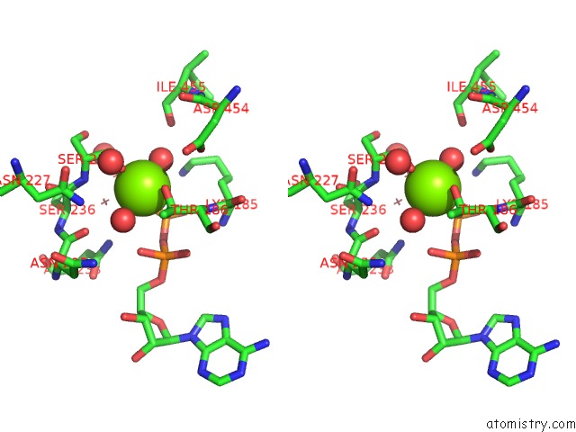
Stereo pair view
Mono view
Stereo pair view
A full contact list of Magnesium with other atoms in the Mg binding
site number 1 of Crystal Structure of Myosin-2 in Complex with Adp-Metavanadate within 5.0Å range:
|
Reference:
R.Fedorov,
M.Bohl,
G.Tsiavaliaris,
F.K.Hartmann,
M.H.Taft,
P.Baruch,
B.Brenner,
R.Martin,
H.Knolker,
H.O.Gutzeit,
D.J.Manstein.
The Mechanism of Pentabromopseudilin Inhibition of Myosin Motor Activity. Nat.Struct.Mol.Biol. V. 16 80 2009.
ISSN: ISSN 1545-9993
PubMed: 19122661
DOI: 10.1038/NSMB.1542
Page generated: Wed Aug 14 00:48:20 2024
ISSN: ISSN 1545-9993
PubMed: 19122661
DOI: 10.1038/NSMB.1542
Last articles
Fe in 2YXOFe in 2YRS
Fe in 2YXC
Fe in 2YNM
Fe in 2YVJ
Fe in 2YP1
Fe in 2YU2
Fe in 2YU1
Fe in 2YQB
Fe in 2YOO