Magnesium »
PDB 2jga-2mse »
2kfz »
Magnesium in PDB 2kfz: Klenow Fragment with Bridging-Sulfur Substrate and Zinc Only
Enzymatic activity of Klenow Fragment with Bridging-Sulfur Substrate and Zinc Only
All present enzymatic activity of Klenow Fragment with Bridging-Sulfur Substrate and Zinc Only:
2.7.7.7;
2.7.7.7;
Protein crystallography data
The structure of Klenow Fragment with Bridging-Sulfur Substrate and Zinc Only, PDB code: 2kfz
was solved by
C.A.Brautigam,
S.Sun,
J.A.Piccirilli,
T.A.Steitz,
with X-Ray Crystallography technique. A brief refinement statistics is given in the table below:
| Resolution Low / High (Å) | 20.00 / 2.03 |
| Space group | P 43 |
| Cell size a, b, c (Å), α, β, γ (°) | 102.900, 102.900, 86.400, 90.00, 90.00, 90.00 |
| R / Rfree (%) | 21.5 / 25.5 |
Other elements in 2kfz:
The structure of Klenow Fragment with Bridging-Sulfur Substrate and Zinc Only also contains other interesting chemical elements:
| Zinc | (Zn) | 4 atoms |
Magnesium Binding Sites:
The binding sites of Magnesium atom in the Klenow Fragment with Bridging-Sulfur Substrate and Zinc Only
(pdb code 2kfz). This binding sites where shown within
5.0 Angstroms radius around Magnesium atom.
In total only one binding site of Magnesium was determined in the Klenow Fragment with Bridging-Sulfur Substrate and Zinc Only, PDB code: 2kfz:
In total only one binding site of Magnesium was determined in the Klenow Fragment with Bridging-Sulfur Substrate and Zinc Only, PDB code: 2kfz:
Magnesium binding site 1 out of 1 in 2kfz
Go back to
Magnesium binding site 1 out
of 1 in the Klenow Fragment with Bridging-Sulfur Substrate and Zinc Only
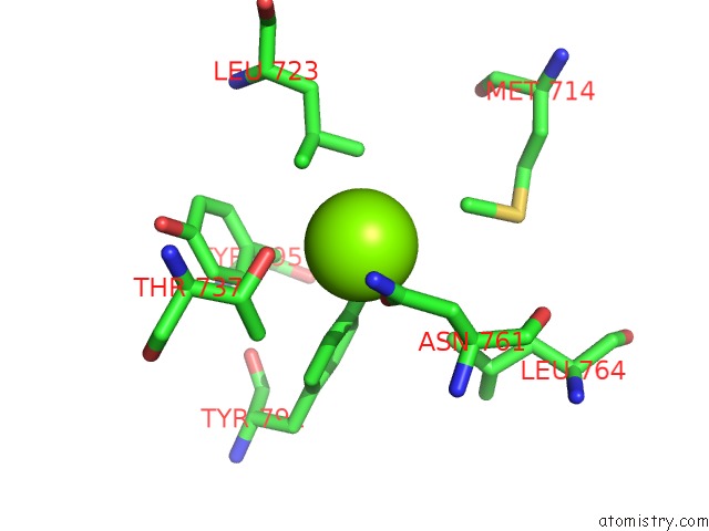
Mono view
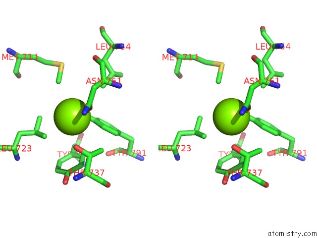
Stereo pair view
Mono view
Stereo pair view
A full contact list of Magnesium with other atoms in the Mg binding
site number 1 of Klenow Fragment with Bridging-Sulfur Substrate and Zinc Only within 5.0Å range:
|
Reference:
C.A.Brautigam,
S.Sun,
J.A.Piccirilli,
T.A.Steitz.
Structures of Normal Single-Stranded Dna and Deoxyribo-3'-S-Phosphorothiolates Bound to the 3'-5' Exonucleolytic Active Site of Dna Polymerase I From Escherichia Coli. Biochemistry V. 38 696 1999.
ISSN: ISSN 0006-2960
PubMed: 9888810
DOI: 10.1021/BI981537G
Page generated: Wed Aug 14 00:50:11 2024
ISSN: ISSN 0006-2960
PubMed: 9888810
DOI: 10.1021/BI981537G
Last articles
Cl in 5VWVCl in 5VVI
Cl in 5VWM
Cl in 5VVB
Cl in 5VVC
Cl in 5VVD
Cl in 5VVE
Cl in 5VUS
Cl in 5VTK
Cl in 5VSV