Magnesium »
PDB 2mtk-2o52 »
2nok »
Magnesium in PDB 2nok: Crystal Structure of An Rna Domain From Hepatitis C Virus.
Protein crystallography data
The structure of Crystal Structure of An Rna Domain From Hepatitis C Virus., PDB code: 2nok
was solved by
S.M.Dibrov,
H.Johnston-Cos,
Y.H.Weng,
with X-Ray Crystallography technique. A brief refinement statistics is given in the table below:
| Resolution Low / High (Å) | 20.00 / 3.00 |
| Space group | P 21 21 21 |
| Cell size a, b, c (Å), α, β, γ (°) | 62.098, 65.351, 92.400, 90.00, 90.00, 90.00 |
| R / Rfree (%) | 25.7 / 30.2 |
Other elements in 2nok:
The structure of Crystal Structure of An Rna Domain From Hepatitis C Virus. also contains other interesting chemical elements:
| Manganese | (Mn) | 5 atoms |
Magnesium Binding Sites:
The binding sites of Magnesium atom in the Crystal Structure of An Rna Domain From Hepatitis C Virus.
(pdb code 2nok). This binding sites where shown within
5.0 Angstroms radius around Magnesium atom.
In total 8 binding sites of Magnesium where determined in the Crystal Structure of An Rna Domain From Hepatitis C Virus., PDB code: 2nok:
Jump to Magnesium binding site number: 1; 2; 3; 4; 5; 6; 7; 8;
In total 8 binding sites of Magnesium where determined in the Crystal Structure of An Rna Domain From Hepatitis C Virus., PDB code: 2nok:
Jump to Magnesium binding site number: 1; 2; 3; 4; 5; 6; 7; 8;
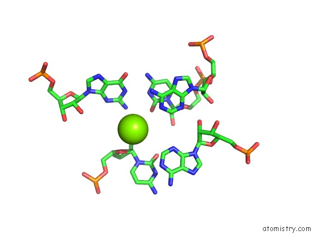
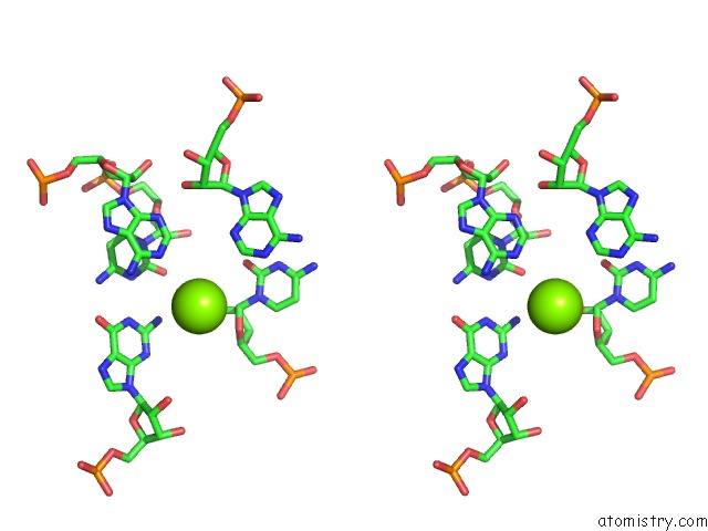
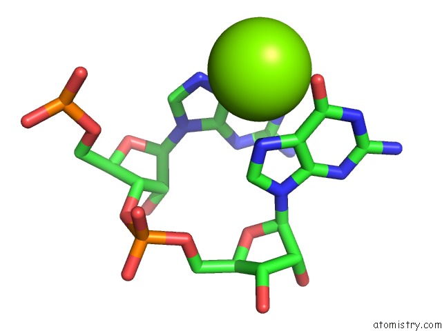
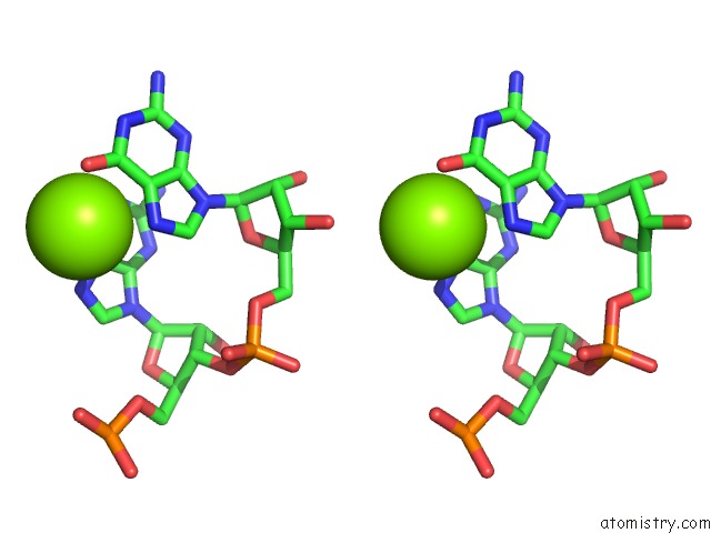
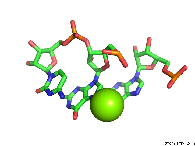
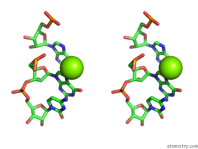
Magnesium binding site 1 out of 8 in 2nok
Go back to
Magnesium binding site 1 out
of 8 in the Crystal Structure of An Rna Domain From Hepatitis C Virus.
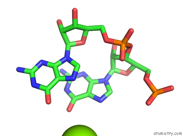
Mono view
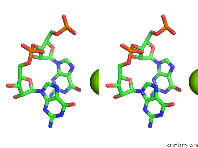
Stereo pair view
Mono view
Stereo pair view
A full contact list of Magnesium with other atoms in the Mg binding
site number 1 of Crystal Structure of An Rna Domain From Hepatitis C Virus. within 5.0Å range:
|
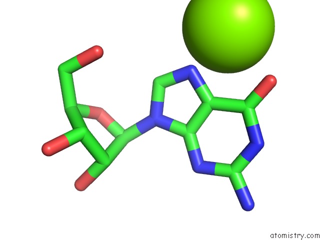
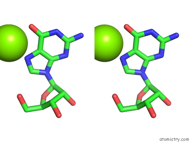
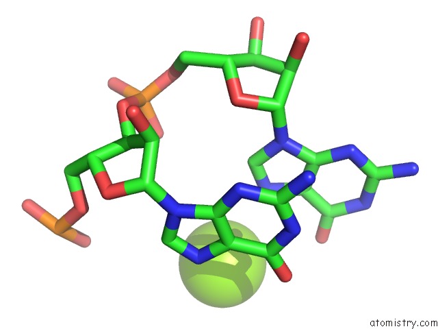
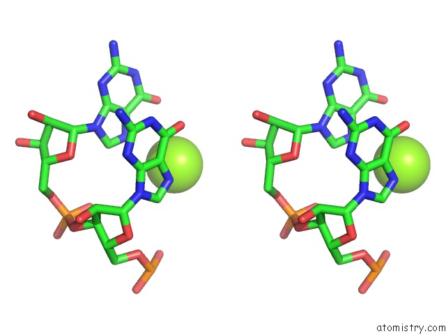
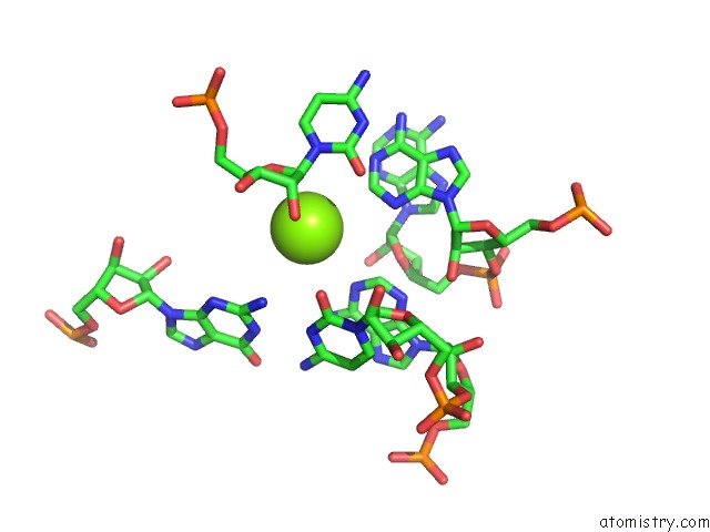
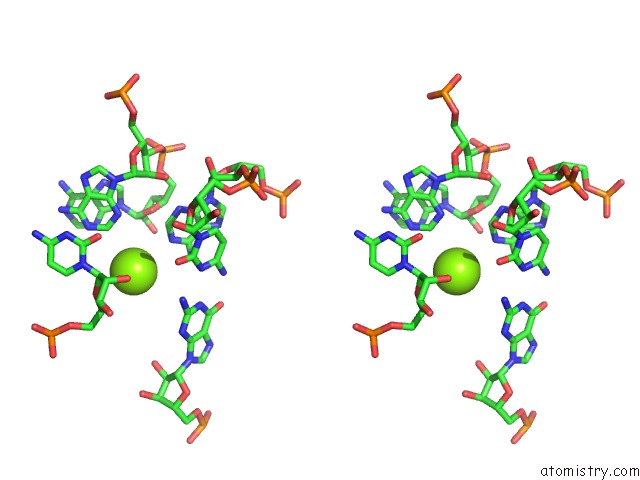
Magnesium binding site 2 out of 8 in 2nok
Go back to
Magnesium binding site 2 out
of 8 in the Crystal Structure of An Rna Domain From Hepatitis C Virus.
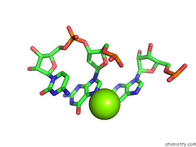
Mono view
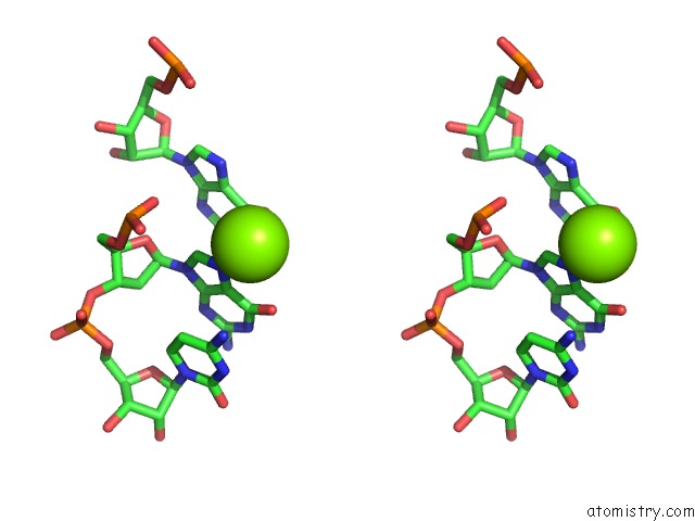
Stereo pair view
Mono view
Stereo pair view
A full contact list of Magnesium with other atoms in the Mg binding
site number 2 of Crystal Structure of An Rna Domain From Hepatitis C Virus. within 5.0Å range:
|
Magnesium binding site 3 out of 8 in 2nok
Go back to
Magnesium binding site 3 out
of 8 in the Crystal Structure of An Rna Domain From Hepatitis C Virus.
Mono view
Stereo pair view
Mono view
Stereo pair view
A full contact list of Magnesium with other atoms in the Mg binding
site number 3 of Crystal Structure of An Rna Domain From Hepatitis C Virus. within 5.0Å range:
|
Magnesium binding site 4 out of 8 in 2nok
Go back to
Magnesium binding site 4 out
of 8 in the Crystal Structure of An Rna Domain From Hepatitis C Virus.
Mono view
Stereo pair view
Mono view
Stereo pair view
A full contact list of Magnesium with other atoms in the Mg binding
site number 4 of Crystal Structure of An Rna Domain From Hepatitis C Virus. within 5.0Å range:
|
Magnesium binding site 5 out of 8 in 2nok
Go back to
Magnesium binding site 5 out
of 8 in the Crystal Structure of An Rna Domain From Hepatitis C Virus.
Mono view
Stereo pair view
Mono view
Stereo pair view
A full contact list of Magnesium with other atoms in the Mg binding
site number 5 of Crystal Structure of An Rna Domain From Hepatitis C Virus. within 5.0Å range:
|
Magnesium binding site 6 out of 8 in 2nok
Go back to
Magnesium binding site 6 out
of 8 in the Crystal Structure of An Rna Domain From Hepatitis C Virus.
Mono view
Stereo pair view
Mono view
Stereo pair view
A full contact list of Magnesium with other atoms in the Mg binding
site number 6 of Crystal Structure of An Rna Domain From Hepatitis C Virus. within 5.0Å range:
|
Magnesium binding site 7 out of 8 in 2nok
Go back to
Magnesium binding site 7 out
of 8 in the Crystal Structure of An Rna Domain From Hepatitis C Virus.
Mono view
Stereo pair view
Mono view
Stereo pair view
A full contact list of Magnesium with other atoms in the Mg binding
site number 7 of Crystal Structure of An Rna Domain From Hepatitis C Virus. within 5.0Å range:
|
Magnesium binding site 8 out of 8 in 2nok
Go back to
Magnesium binding site 8 out
of 8 in the Crystal Structure of An Rna Domain From Hepatitis C Virus.
Mono view
Stereo pair view
Mono view
Stereo pair view
A full contact list of Magnesium with other atoms in the Mg binding
site number 8 of Crystal Structure of An Rna Domain From Hepatitis C Virus. within 5.0Å range:
|
Reference:
S.M.Dibrov,
H.Johnston-Cox,
Y.H.Weng,
T.Hermann.
Functional Architecture of Hcv Ires Domain II Stabilized By Divalent Metal Ions in the Crystal and in Solution. Angew.Chem.Int.Ed.Engl. V. 46 226 2007.
ISSN: ESSN 0570-0833
PubMed: 17131443
DOI: 10.1002/ANIE.200603807
Page generated: Wed Aug 14 00:53:34 2024
ISSN: ESSN 0570-0833
PubMed: 17131443
DOI: 10.1002/ANIE.200603807
Last articles
Zn in 9MJ5Zn in 9HNW
Zn in 9G0L
Zn in 9FNE
Zn in 9DZN
Zn in 9E0I
Zn in 9D32
Zn in 9DAK
Zn in 8ZXC
Zn in 8ZUF