Magnesium »
PDB 2o56-2oh6 »
2ode »
Magnesium in PDB 2ode: Crystal Structure of the Heterodimeric Complex of Human RGS8 and Activated Gi Alpha 3
Protein crystallography data
The structure of Crystal Structure of the Heterodimeric Complex of Human RGS8 and Activated Gi Alpha 3, PDB code: 2ode
was solved by
C.Gileadi,
M.Soundararajan,
A.P.Turnbull,
J.M.Elkins,
E.Papagrigoriou,
A.C.W.Pike,
G.Bunkoczi,
F.Gorrec,
C.Umeano,
F.Von Delft,
J.Weigelt,
A.Edwards,
C.H.Arrowsmith,
M.Sundstrom,
D.A.Doyle,
Structural Genomicsconsortium (Sgc),
with X-Ray Crystallography technique. A brief refinement statistics is given in the table below:
| Resolution Low / High (Å) | 29.40 / 1.90 |
| Space group | P 21 21 2 |
| Cell size a, b, c (Å), α, β, γ (°) | 112.837, 130.216, 68.519, 90.00, 90.00, 90.00 |
| R / Rfree (%) | 18 / 21.1 |
Other elements in 2ode:
The structure of Crystal Structure of the Heterodimeric Complex of Human RGS8 and Activated Gi Alpha 3 also contains other interesting chemical elements:
| Fluorine | (F) | 8 atoms |
| Aluminium | (Al) | 2 atoms |
Magnesium Binding Sites:
The binding sites of Magnesium atom in the Crystal Structure of the Heterodimeric Complex of Human RGS8 and Activated Gi Alpha 3
(pdb code 2ode). This binding sites where shown within
5.0 Angstroms radius around Magnesium atom.
In total 2 binding sites of Magnesium where determined in the Crystal Structure of the Heterodimeric Complex of Human RGS8 and Activated Gi Alpha 3, PDB code: 2ode:
Jump to Magnesium binding site number: 1; 2;
In total 2 binding sites of Magnesium where determined in the Crystal Structure of the Heterodimeric Complex of Human RGS8 and Activated Gi Alpha 3, PDB code: 2ode:
Jump to Magnesium binding site number: 1; 2;
Magnesium binding site 1 out of 2 in 2ode
Go back to
Magnesium binding site 1 out
of 2 in the Crystal Structure of the Heterodimeric Complex of Human RGS8 and Activated Gi Alpha 3
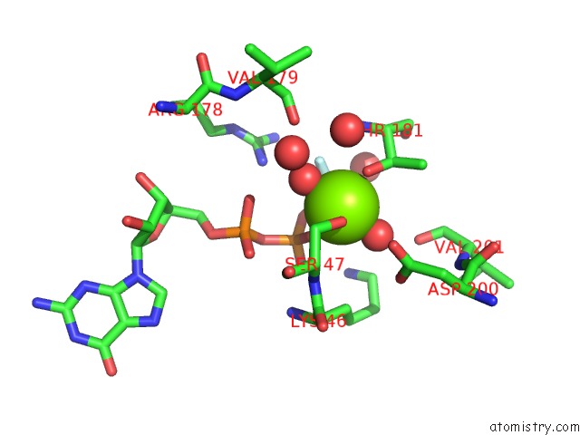
Mono view
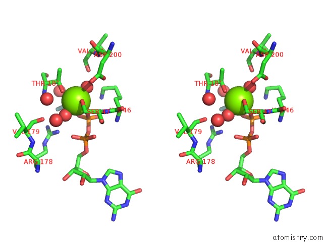
Stereo pair view
Mono view
Stereo pair view
A full contact list of Magnesium with other atoms in the Mg binding
site number 1 of Crystal Structure of the Heterodimeric Complex of Human RGS8 and Activated Gi Alpha 3 within 5.0Å range:
|
Magnesium binding site 2 out of 2 in 2ode
Go back to
Magnesium binding site 2 out
of 2 in the Crystal Structure of the Heterodimeric Complex of Human RGS8 and Activated Gi Alpha 3
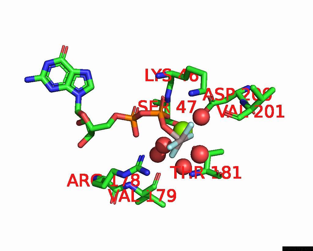
Mono view
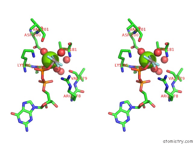
Stereo pair view
Mono view
Stereo pair view
A full contact list of Magnesium with other atoms in the Mg binding
site number 2 of Crystal Structure of the Heterodimeric Complex of Human RGS8 and Activated Gi Alpha 3 within 5.0Å range:
|
Reference:
M.Soundararajan,
F.S.Willard,
A.J.Kimple,
A.P.Turnbull,
L.J.Ball,
G.A.Schoch,
C.Gileadi,
O.Y.Fedorov,
E.F.Dowler,
V.A.Higman,
S.Q.Hutsell,
M.Sundstrom,
D.A.Doyle,
D.P.Siderovski.
Structural Diversity in the Rgs Domain and Its Interaction with Heterotrimeric G Protein Alpha-Subunits. Proc.Natl.Acad.Sci.Usa V. 105 6457 2008.
ISSN: ISSN 0027-8424
PubMed: 18434541
DOI: 10.1073/PNAS.0801508105
Page generated: Sun Aug 10 12:25:52 2025
ISSN: ISSN 0027-8424
PubMed: 18434541
DOI: 10.1073/PNAS.0801508105
Last articles
Mg in 6CA4Mg in 6C90
Mg in 6CA0
Mg in 6C9Y
Mg in 6C8Z
Mg in 6C8P
Mg in 6C8N
Mg in 6C8O
Mg in 6C8D
Mg in 6C8L