Magnesium »
PDB 2o56-2oh6 »
2ofw »
Magnesium in PDB 2ofw: Crystal Structure of the Apsk Domain of Human PAPSS1 Complexed with 2 Aps Molecules
Enzymatic activity of Crystal Structure of the Apsk Domain of Human PAPSS1 Complexed with 2 Aps Molecules
All present enzymatic activity of Crystal Structure of the Apsk Domain of Human PAPSS1 Complexed with 2 Aps Molecules:
2.7.1.25;
2.7.1.25;
Protein crystallography data
The structure of Crystal Structure of the Apsk Domain of Human PAPSS1 Complexed with 2 Aps Molecules, PDB code: 2ofw
was solved by
N.Sekulic,
A.Lavie,
with X-Ray Crystallography technique. A brief refinement statistics is given in the table below:
| Resolution Low / High (Å) | 10.00 / 2.05 |
| Space group | C 1 2 1 |
| Cell size a, b, c (Å), α, β, γ (°) | 181.190, 69.030, 150.610, 90.00, 116.61, 90.00 |
| R / Rfree (%) | 21.7 / 28.2 |
Magnesium Binding Sites:
The binding sites of Magnesium atom in the Crystal Structure of the Apsk Domain of Human PAPSS1 Complexed with 2 Aps Molecules
(pdb code 2ofw). This binding sites where shown within
5.0 Angstroms radius around Magnesium atom.
In total 6 binding sites of Magnesium where determined in the Crystal Structure of the Apsk Domain of Human PAPSS1 Complexed with 2 Aps Molecules, PDB code: 2ofw:
Jump to Magnesium binding site number: 1; 2; 3; 4; 5; 6;
In total 6 binding sites of Magnesium where determined in the Crystal Structure of the Apsk Domain of Human PAPSS1 Complexed with 2 Aps Molecules, PDB code: 2ofw:
Jump to Magnesium binding site number: 1; 2; 3; 4; 5; 6;
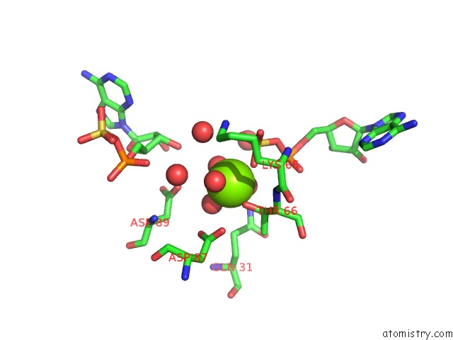
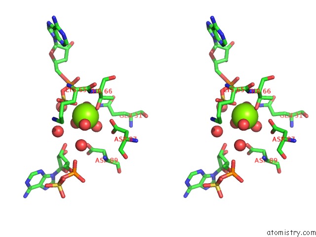
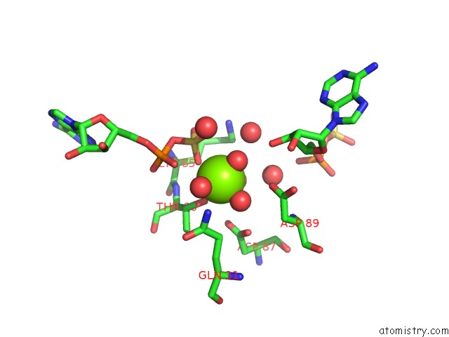
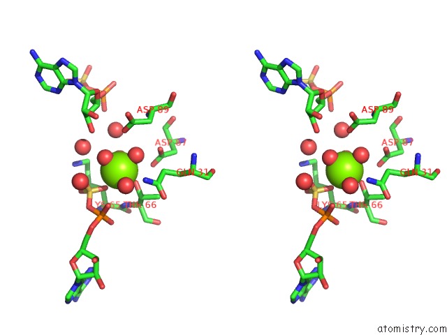
Magnesium binding site 1 out of 6 in 2ofw
Go back to
Magnesium binding site 1 out
of 6 in the Crystal Structure of the Apsk Domain of Human PAPSS1 Complexed with 2 Aps Molecules
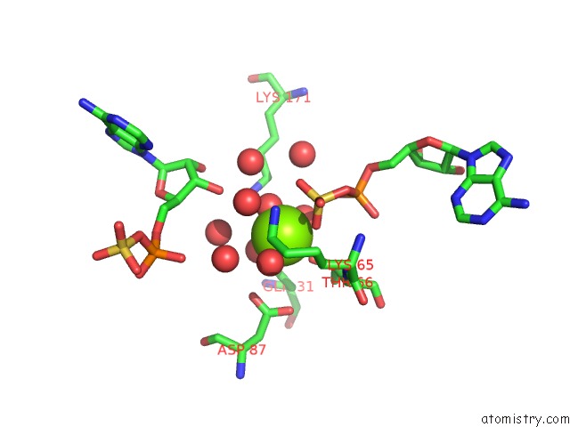
Mono view
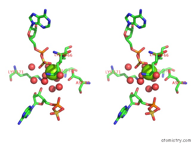
Stereo pair view
Mono view
Stereo pair view
A full contact list of Magnesium with other atoms in the Mg binding
site number 1 of Crystal Structure of the Apsk Domain of Human PAPSS1 Complexed with 2 Aps Molecules within 5.0Å range:
|
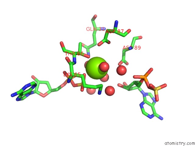
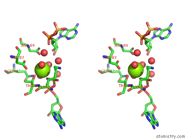
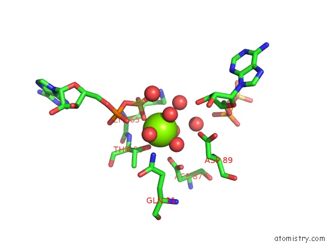
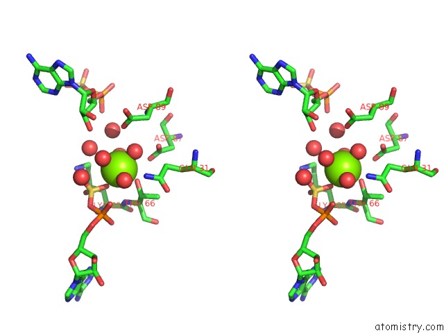
Magnesium binding site 2 out of 6 in 2ofw
Go back to
Magnesium binding site 2 out
of 6 in the Crystal Structure of the Apsk Domain of Human PAPSS1 Complexed with 2 Aps Molecules
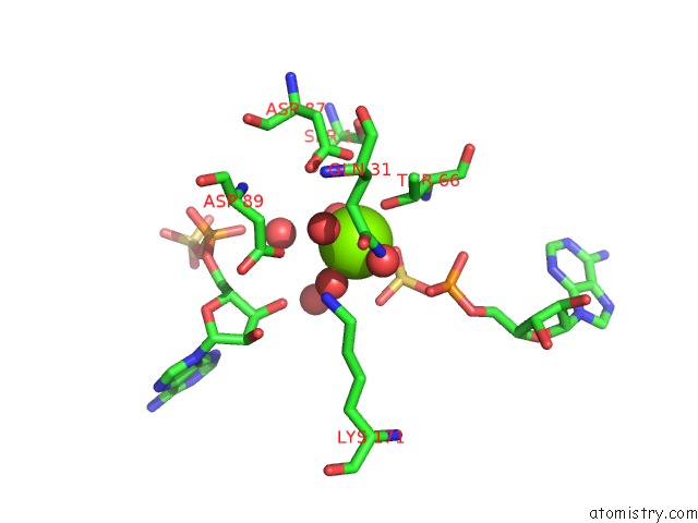
Mono view
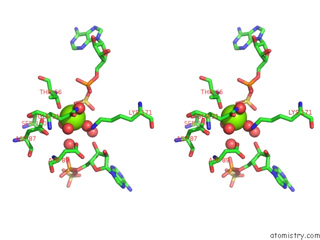
Stereo pair view
Mono view
Stereo pair view
A full contact list of Magnesium with other atoms in the Mg binding
site number 2 of Crystal Structure of the Apsk Domain of Human PAPSS1 Complexed with 2 Aps Molecules within 5.0Å range:
|
Magnesium binding site 3 out of 6 in 2ofw
Go back to
Magnesium binding site 3 out
of 6 in the Crystal Structure of the Apsk Domain of Human PAPSS1 Complexed with 2 Aps Molecules
Mono view
Stereo pair view
Mono view
Stereo pair view
A full contact list of Magnesium with other atoms in the Mg binding
site number 3 of Crystal Structure of the Apsk Domain of Human PAPSS1 Complexed with 2 Aps Molecules within 5.0Å range:
|
Magnesium binding site 4 out of 6 in 2ofw
Go back to
Magnesium binding site 4 out
of 6 in the Crystal Structure of the Apsk Domain of Human PAPSS1 Complexed with 2 Aps Molecules
Mono view
Stereo pair view
Mono view
Stereo pair view
A full contact list of Magnesium with other atoms in the Mg binding
site number 4 of Crystal Structure of the Apsk Domain of Human PAPSS1 Complexed with 2 Aps Molecules within 5.0Å range:
|
Magnesium binding site 5 out of 6 in 2ofw
Go back to
Magnesium binding site 5 out
of 6 in the Crystal Structure of the Apsk Domain of Human PAPSS1 Complexed with 2 Aps Molecules
Mono view
Stereo pair view
Mono view
Stereo pair view
A full contact list of Magnesium with other atoms in the Mg binding
site number 5 of Crystal Structure of the Apsk Domain of Human PAPSS1 Complexed with 2 Aps Molecules within 5.0Å range:
|
Magnesium binding site 6 out of 6 in 2ofw
Go back to
Magnesium binding site 6 out
of 6 in the Crystal Structure of the Apsk Domain of Human PAPSS1 Complexed with 2 Aps Molecules
Mono view
Stereo pair view
Mono view
Stereo pair view
A full contact list of Magnesium with other atoms in the Mg binding
site number 6 of Crystal Structure of the Apsk Domain of Human PAPSS1 Complexed with 2 Aps Molecules within 5.0Å range:
|
Reference:
N.Sekulic,
K.Dietrich,
I.Paarmann,
S.Ort,
M.Konrad,
A.Lavie.
Elucidation of the Active Conformation of the Aps-Kinase Domain of Human Paps Synthetase 1. J.Mol.Biol. V. 367 488 2007.
ISSN: ISSN 0022-2836
PubMed: 17276460
DOI: 10.1016/J.JMB.2007.01.025
Page generated: Wed Aug 14 01:26:59 2024
ISSN: ISSN 0022-2836
PubMed: 17276460
DOI: 10.1016/J.JMB.2007.01.025
Last articles
Zn in 9MJ5Zn in 9HNW
Zn in 9G0L
Zn in 9FNE
Zn in 9DZN
Zn in 9E0I
Zn in 9D32
Zn in 9DAK
Zn in 8ZXC
Zn in 8ZUF