Magnesium »
PDB 2oh7-2oup »
2oi7 »
Magnesium in PDB 2oi7: E. Coli Glmu- Complex with Udp-Glcnac, Desulpho-Coa and Glcnac-1-PO4
Enzymatic activity of E. Coli Glmu- Complex with Udp-Glcnac, Desulpho-Coa and Glcnac-1-PO4
All present enzymatic activity of E. Coli Glmu- Complex with Udp-Glcnac, Desulpho-Coa and Glcnac-1-PO4:
2.3.1.157; 2.7.7.23;
2.3.1.157; 2.7.7.23;
Protein crystallography data
The structure of E. Coli Glmu- Complex with Udp-Glcnac, Desulpho-Coa and Glcnac-1-PO4, PDB code: 2oi7
was solved by
L.R.Olsen,
M.W.Vetting,
S.L.Roderick,
with X-Ray Crystallography technique. A brief refinement statistics is given in the table below:
| Resolution Low / High (Å) | 27.45 / 2.54 |
| Space group | H 3 2 |
| Cell size a, b, c (Å), α, β, γ (°) | 102.969, 102.969, 644.076, 90.00, 90.00, 120.00 |
| R / Rfree (%) | 20.9 / 24.7 |
Other elements in 2oi7:
The structure of E. Coli Glmu- Complex with Udp-Glcnac, Desulpho-Coa and Glcnac-1-PO4 also contains other interesting chemical elements:
| Cobalt | (Co) | 3 atoms |
Magnesium Binding Sites:
The binding sites of Magnesium atom in the E. Coli Glmu- Complex with Udp-Glcnac, Desulpho-Coa and Glcnac-1-PO4
(pdb code 2oi7). This binding sites where shown within
5.0 Angstroms radius around Magnesium atom.
In total only one binding site of Magnesium was determined in the E. Coli Glmu- Complex with Udp-Glcnac, Desulpho-Coa and Glcnac-1-PO4, PDB code: 2oi7:
In total only one binding site of Magnesium was determined in the E. Coli Glmu- Complex with Udp-Glcnac, Desulpho-Coa and Glcnac-1-PO4, PDB code: 2oi7:
Magnesium binding site 1 out of 1 in 2oi7
Go back to
Magnesium binding site 1 out
of 1 in the E. Coli Glmu- Complex with Udp-Glcnac, Desulpho-Coa and Glcnac-1-PO4
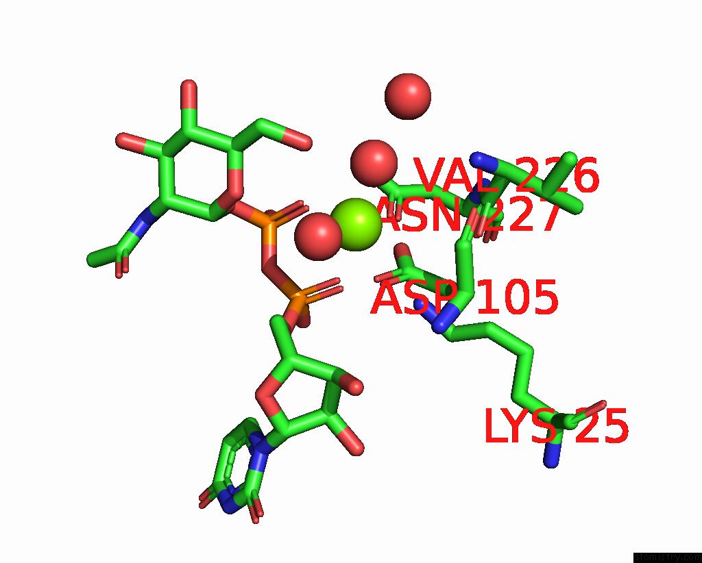
Mono view
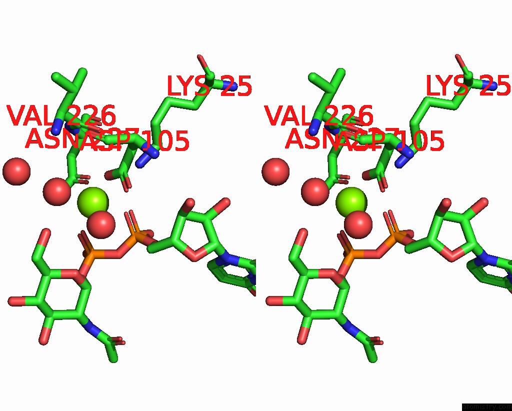
Stereo pair view
Mono view
Stereo pair view
A full contact list of Magnesium with other atoms in the Mg binding
site number 1 of E. Coli Glmu- Complex with Udp-Glcnac, Desulpho-Coa and Glcnac-1-PO4 within 5.0Å range:
|
Reference:
L.R.Olsen,
M.W.Vetting,
S.L.Roderick.
Structure of the E. Coli Bifunctional Glmu Acetyltransferase Active Site with Substrates and Products. Protein Sci. V. 16 1230 2007.
ISSN: ISSN 0961-8368
PubMed: 17473010
DOI: 10.1110/PS.072779707
Page generated: Sun Aug 10 12:28:02 2025
ISSN: ISSN 0961-8368
PubMed: 17473010
DOI: 10.1110/PS.072779707
Last articles
Mg in 6IAHMg in 6IB7
Mg in 6IAN
Mg in 6I9P
Mg in 6I9T
Mg in 6IAF
Mg in 6IAE
Mg in 6IA7
Mg in 6I8F
Mg in 6I5X