Magnesium »
PDB 2oh7-2oup »
2oiu »
Magnesium in PDB 2oiu: L1 Ribozyme Ligase Circular Adduct
Protein crystallography data
The structure of L1 Ribozyme Ligase Circular Adduct, PDB code: 2oiu
was solved by
M.P.Robertson,
W.G.Scott,
with X-Ray Crystallography technique. A brief refinement statistics is given in the table below:
| Resolution Low / High (Å) | 31.34 / 2.60 |
| Space group | P 1 21 1 |
| Cell size a, b, c (Å), α, β, γ (°) | 45.290, 100.018, 71.930, 90.00, 104.42, 90.00 |
| R / Rfree (%) | 20.3 / 23.8 |
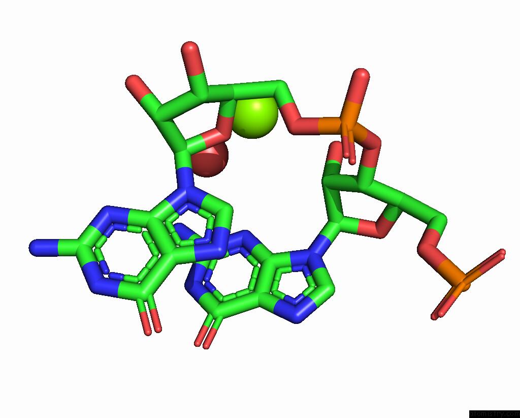
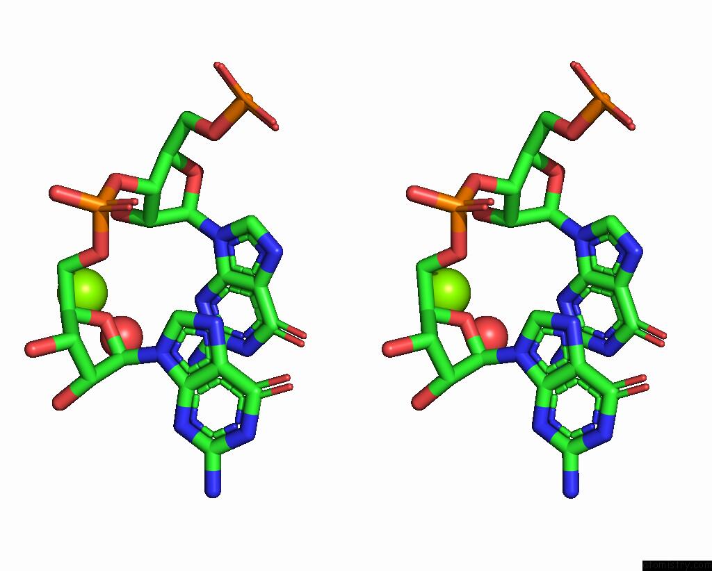
Magnesium Binding Sites:
The binding sites of Magnesium atom in the L1 Ribozyme Ligase Circular Adduct
(pdb code 2oiu). This binding sites where shown within
5.0 Angstroms radius around Magnesium atom.
In total 7 binding sites of Magnesium where determined in the L1 Ribozyme Ligase Circular Adduct, PDB code: 2oiu:
Jump to Magnesium binding site number: 1; 2; 3; 4; 5; 6; 7;
In total 7 binding sites of Magnesium where determined in the L1 Ribozyme Ligase Circular Adduct, PDB code: 2oiu:
Jump to Magnesium binding site number: 1; 2; 3; 4; 5; 6; 7;
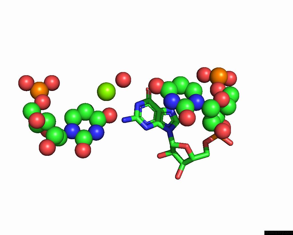
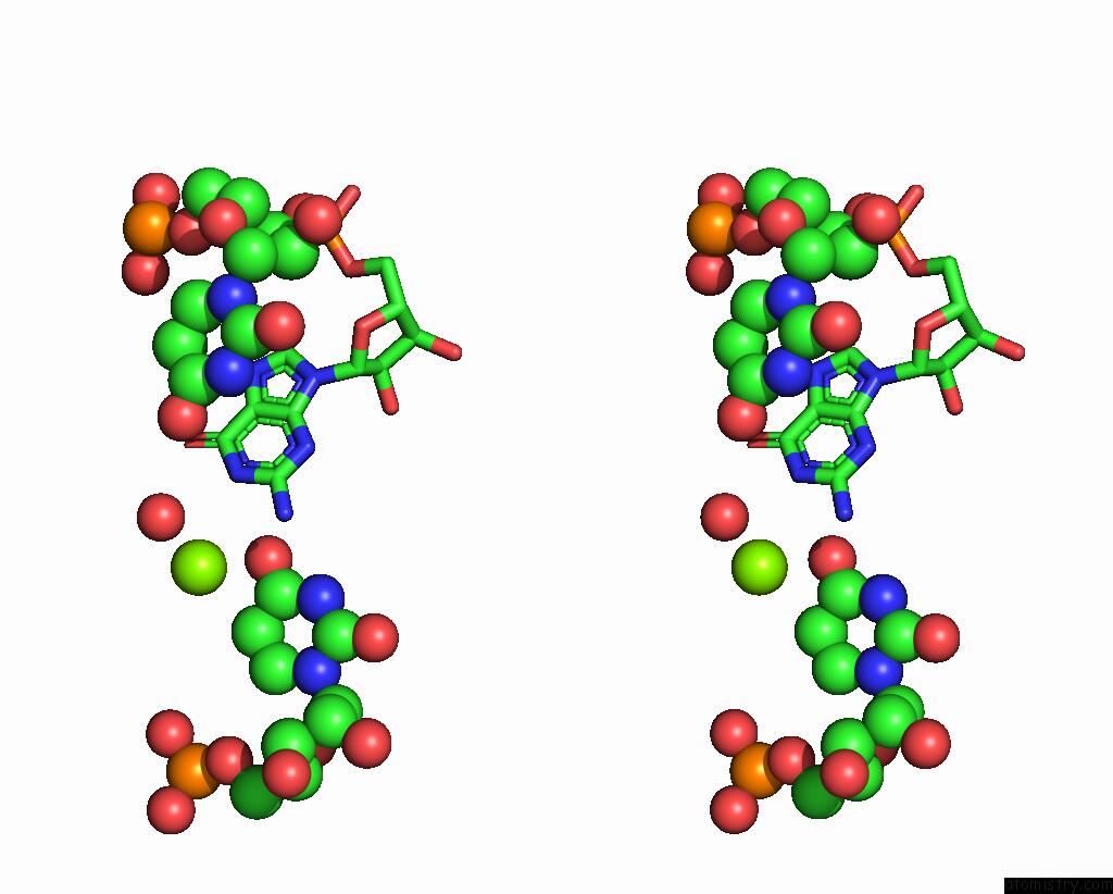
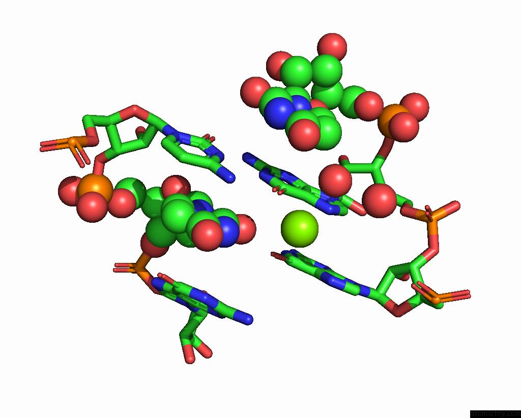
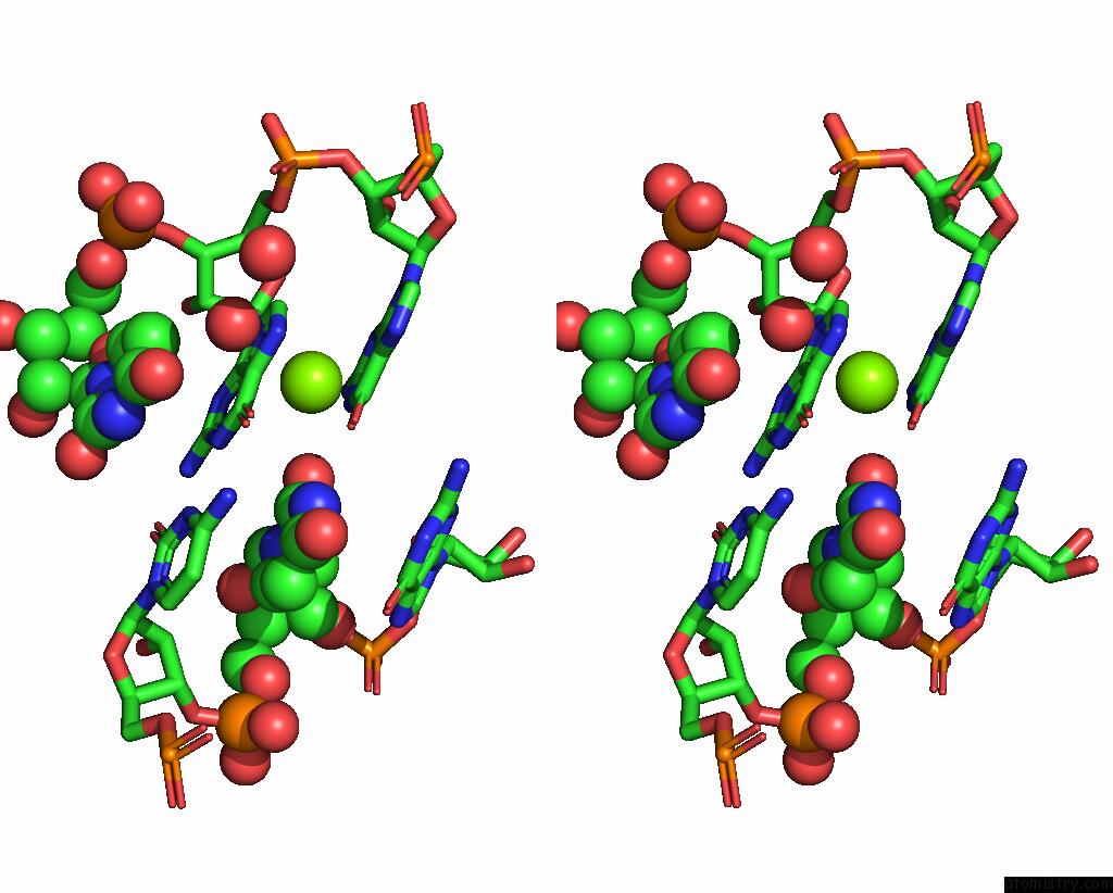
Magnesium binding site 1 out of 7 in 2oiu
Go back to
Magnesium binding site 1 out
of 7 in the L1 Ribozyme Ligase Circular Adduct
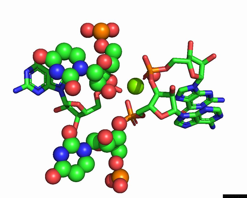
Mono view
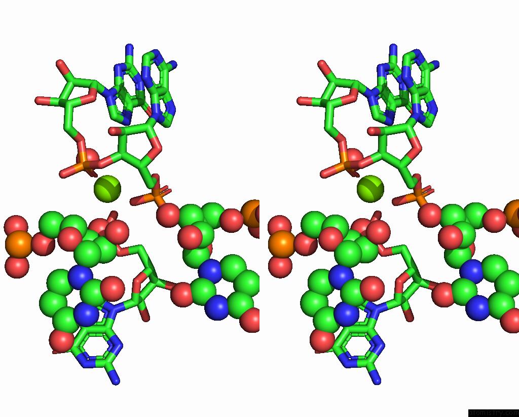
Stereo pair view
Mono view
Stereo pair view
A full contact list of Magnesium with other atoms in the Mg binding
site number 1 of L1 Ribozyme Ligase Circular Adduct within 5.0Å range:
|
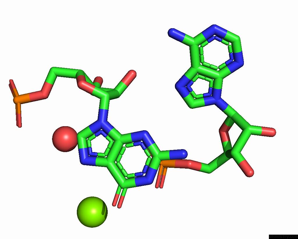
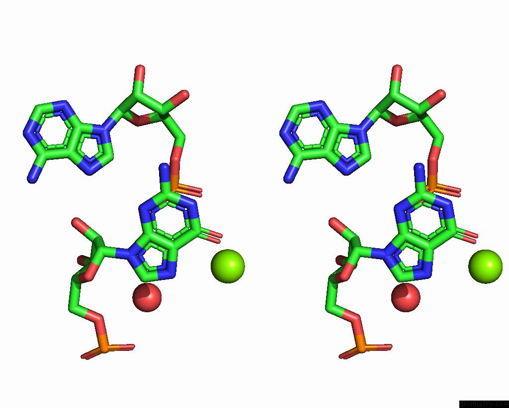
Magnesium binding site 2 out of 7 in 2oiu
Go back to
Magnesium binding site 2 out
of 7 in the L1 Ribozyme Ligase Circular Adduct
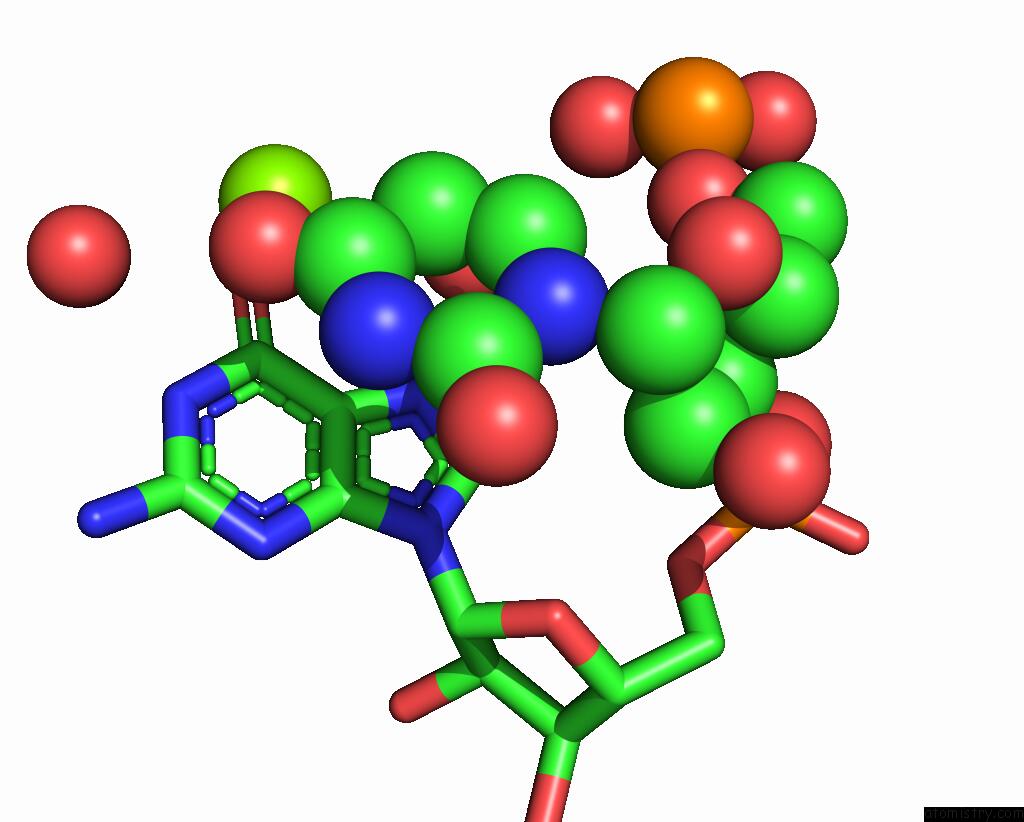
Mono view
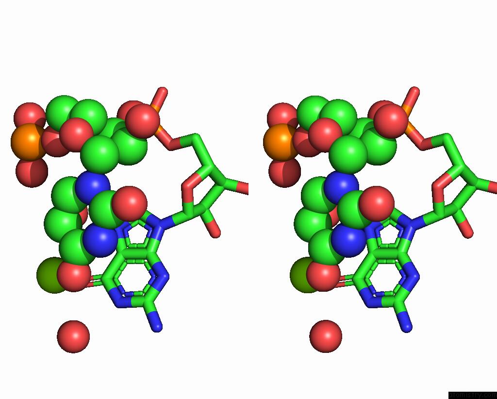
Stereo pair view
Mono view
Stereo pair view
A full contact list of Magnesium with other atoms in the Mg binding
site number 2 of L1 Ribozyme Ligase Circular Adduct within 5.0Å range:
|
Magnesium binding site 3 out of 7 in 2oiu
Go back to
Magnesium binding site 3 out
of 7 in the L1 Ribozyme Ligase Circular Adduct
Mono view
Stereo pair view
Mono view
Stereo pair view
A full contact list of Magnesium with other atoms in the Mg binding
site number 3 of L1 Ribozyme Ligase Circular Adduct within 5.0Å range:
|
Magnesium binding site 4 out of 7 in 2oiu
Go back to
Magnesium binding site 4 out
of 7 in the L1 Ribozyme Ligase Circular Adduct
Mono view
Stereo pair view
Mono view
Stereo pair view
A full contact list of Magnesium with other atoms in the Mg binding
site number 4 of L1 Ribozyme Ligase Circular Adduct within 5.0Å range:
|
Magnesium binding site 5 out of 7 in 2oiu
Go back to
Magnesium binding site 5 out
of 7 in the L1 Ribozyme Ligase Circular Adduct
Mono view
Stereo pair view
Mono view
Stereo pair view
| A full contact list of Magnesium with other atoms in the Mg binding site number 5 of L1 Ribozyme Ligase Circular Adduct within 5.0Å range: |
Magnesium binding site 6 out of 7 in 2oiu
Go back to
Magnesium binding site 6 out
of 7 in the L1 Ribozyme Ligase Circular Adduct
Mono view
Stereo pair view
Mono view
Stereo pair view
A full contact list of Magnesium with other atoms in the Mg binding
site number 6 of L1 Ribozyme Ligase Circular Adduct within 5.0Å range:
|
Magnesium binding site 7 out of 7 in 2oiu
Go back to
Magnesium binding site 7 out
of 7 in the L1 Ribozyme Ligase Circular Adduct
Mono view
Stereo pair view
Mono view
Stereo pair view
A full contact list of Magnesium with other atoms in the Mg binding
site number 7 of L1 Ribozyme Ligase Circular Adduct within 5.0Å range:
|
Reference:
M.P.Robertson,
W.G.Scott.
The Structural Basis of Ribozyme-Catalyzed Rna Assembly. Science V. 315 1549 2007.
ISSN: ISSN 0036-8075
PubMed: 17363667
DOI: 10.1126/SCIENCE.1136231
Page generated: Sun Aug 10 12:28:15 2025
ISSN: ISSN 0036-8075
PubMed: 17363667
DOI: 10.1126/SCIENCE.1136231
Last articles
Mg in 6CA4Mg in 6C90
Mg in 6CA0
Mg in 6C9Y
Mg in 6C8Z
Mg in 6C8P
Mg in 6C8N
Mg in 6C8O
Mg in 6C8D
Mg in 6C8L