Magnesium »
PDB 2oh7-2oup »
2oke »
Magnesium in PDB 2oke: High Resolution Crystal Structures of Vaccinia Virus Dutpase
Enzymatic activity of High Resolution Crystal Structures of Vaccinia Virus Dutpase
All present enzymatic activity of High Resolution Crystal Structures of Vaccinia Virus Dutpase:
3.6.1.23;
3.6.1.23;
Protein crystallography data
The structure of High Resolution Crystal Structures of Vaccinia Virus Dutpase, PDB code: 2oke
was solved by
N.Schormann,
D.Chattopadhyay,
with X-Ray Crystallography technique. A brief refinement statistics is given in the table below:
| Resolution Low / High (Å) | 13.47 / 2.50 |
| Space group | P 65 |
| Cell size a, b, c (Å), α, β, γ (°) | 120.460, 120.460, 50.100, 90.00, 90.00, 120.00 |
| R / Rfree (%) | 22 / 27.4 |
Other elements in 2oke:
The structure of High Resolution Crystal Structures of Vaccinia Virus Dutpase also contains other interesting chemical elements:
| Chlorine | (Cl) | 1 atom |
Magnesium Binding Sites:
The binding sites of Magnesium atom in the High Resolution Crystal Structures of Vaccinia Virus Dutpase
(pdb code 2oke). This binding sites where shown within
5.0 Angstroms radius around Magnesium atom.
In total only one binding site of Magnesium was determined in the High Resolution Crystal Structures of Vaccinia Virus Dutpase, PDB code: 2oke:
In total only one binding site of Magnesium was determined in the High Resolution Crystal Structures of Vaccinia Virus Dutpase, PDB code: 2oke:
Magnesium binding site 1 out of 1 in 2oke
Go back to
Magnesium binding site 1 out
of 1 in the High Resolution Crystal Structures of Vaccinia Virus Dutpase
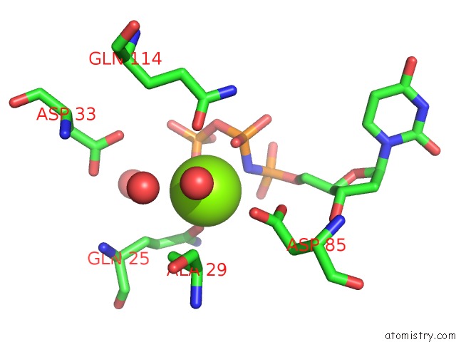
Mono view
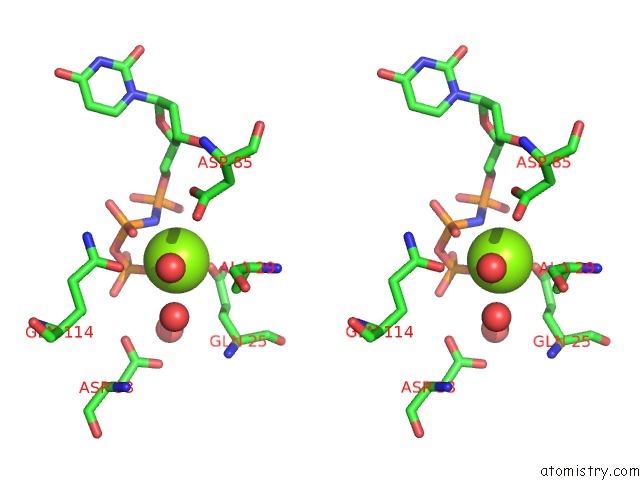
Stereo pair view
Mono view
Stereo pair view
A full contact list of Magnesium with other atoms in the Mg binding
site number 1 of High Resolution Crystal Structures of Vaccinia Virus Dutpase within 5.0Å range:
|
Reference:
A.Samal,
N.Schormann,
W.J.Cook,
L.J.Delucas,
D.Chattopadhyay.
Structures of Vaccinia Virus Dutpase and Its Nucleotide Complexes. Acta Crystallogr.,Sect.D V. 63 571 2007.
ISSN: ISSN 0907-4449
PubMed: 17452782
DOI: 10.1107/S0907444907007871
Page generated: Wed Aug 14 01:30:24 2024
ISSN: ISSN 0907-4449
PubMed: 17452782
DOI: 10.1107/S0907444907007871
Last articles
Zn in 9J0NZn in 9J0O
Zn in 9J0P
Zn in 9FJX
Zn in 9EKB
Zn in 9C0F
Zn in 9CAH
Zn in 9CH0
Zn in 9CH3
Zn in 9CH1