Magnesium »
PDB 2oh7-2oup »
2olr »
Magnesium in PDB 2olr: Crystal Structure of Escherichia Coli Phosphoenolpyruvate Carboxykinase Complexed with Carbon Dioxide, MG2+, Atp
Enzymatic activity of Crystal Structure of Escherichia Coli Phosphoenolpyruvate Carboxykinase Complexed with Carbon Dioxide, MG2+, Atp
All present enzymatic activity of Crystal Structure of Escherichia Coli Phosphoenolpyruvate Carboxykinase Complexed with Carbon Dioxide, MG2+, Atp:
4.1.1.49;
4.1.1.49;
Protein crystallography data
The structure of Crystal Structure of Escherichia Coli Phosphoenolpyruvate Carboxykinase Complexed with Carbon Dioxide, MG2+, Atp, PDB code: 2olr
was solved by
J.J.Cotelesage,
L.T.Delbaere,
H.Goldie,
J.Puttick,
B.Rajabi,
B.Novakovski,
with X-Ray Crystallography technique. A brief refinement statistics is given in the table below:
| Resolution Low / High (Å) | 75.81 / 1.60 |
| Space group | C 1 2 1 |
| Cell size a, b, c (Å), α, β, γ (°) | 124.854, 95.564, 46.476, 90.00, 96.30, 90.00 |
| R / Rfree (%) | 17.5 / 19.8 |
Other elements in 2olr:
The structure of Crystal Structure of Escherichia Coli Phosphoenolpyruvate Carboxykinase Complexed with Carbon Dioxide, MG2+, Atp also contains other interesting chemical elements:
| Chlorine | (Cl) | 1 atom |
Magnesium Binding Sites:
The binding sites of Magnesium atom in the Crystal Structure of Escherichia Coli Phosphoenolpyruvate Carboxykinase Complexed with Carbon Dioxide, MG2+, Atp
(pdb code 2olr). This binding sites where shown within
5.0 Angstroms radius around Magnesium atom.
In total only one binding site of Magnesium was determined in the Crystal Structure of Escherichia Coli Phosphoenolpyruvate Carboxykinase Complexed with Carbon Dioxide, MG2+, Atp, PDB code: 2olr:
In total only one binding site of Magnesium was determined in the Crystal Structure of Escherichia Coli Phosphoenolpyruvate Carboxykinase Complexed with Carbon Dioxide, MG2+, Atp, PDB code: 2olr:
Magnesium binding site 1 out of 1 in 2olr
Go back to
Magnesium binding site 1 out
of 1 in the Crystal Structure of Escherichia Coli Phosphoenolpyruvate Carboxykinase Complexed with Carbon Dioxide, MG2+, Atp
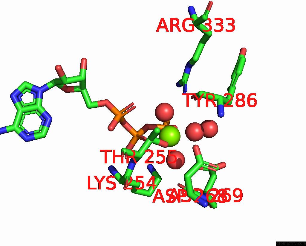
Mono view
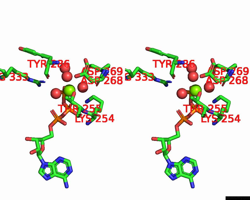
Stereo pair view
Mono view
Stereo pair view
A full contact list of Magnesium with other atoms in the Mg binding
site number 1 of Crystal Structure of Escherichia Coli Phosphoenolpyruvate Carboxykinase Complexed with Carbon Dioxide, MG2+, Atp within 5.0Å range:
|
Reference:
J.J.Cotelesage,
J.Puttick,
H.Goldie,
B.Rajabi,
B.Novakovski,
L.T.Delbaere.
How Does An Enzyme Recognize CO2? Int.J.Biochem.Cell Biol. V. 39 1204 2007.
ISSN: ISSN 1357-2725
PubMed: 17475535
DOI: 10.1016/J.BIOCEL.2007.03.015
Page generated: Sun Aug 10 12:29:31 2025
ISSN: ISSN 1357-2725
PubMed: 17475535
DOI: 10.1016/J.BIOCEL.2007.03.015
Last articles
Mg in 6JNXMg in 6JMG
Mg in 6JNL
Mg in 6JLV
Mg in 6JLN
Mg in 6JLL
Mg in 6JLK
Mg in 6JLM
Mg in 6JLJ
Mg in 6JIL