Magnesium »
PDB 2oh7-2oup »
2ons »
Magnesium in PDB 2ons: Crystal Structure of A. Fulgidus Periplasmic Binding Protein Moda with Bound Tungstate
Protein crystallography data
The structure of Crystal Structure of A. Fulgidus Periplasmic Binding Protein Moda with Bound Tungstate, PDB code: 2ons
was solved by
K.Hollenstein,
D.C.Frei,
K.P.Locher,
with X-Ray Crystallography technique. A brief refinement statistics is given in the table below:
| Resolution Low / High (Å) | 19.79 / 1.55 |
| Space group | P 41 21 2 |
| Cell size a, b, c (Å), α, β, γ (°) | 75.914, 75.914, 115.711, 90.00, 90.00, 90.00 |
| R / Rfree (%) | 18.6 / 20.1 |
Other elements in 2ons:
The structure of Crystal Structure of A. Fulgidus Periplasmic Binding Protein Moda with Bound Tungstate also contains other interesting chemical elements:
| Tungsten | (W) | 1 atom |
Magnesium Binding Sites:
The binding sites of Magnesium atom in the Crystal Structure of A. Fulgidus Periplasmic Binding Protein Moda with Bound Tungstate
(pdb code 2ons). This binding sites where shown within
5.0 Angstroms radius around Magnesium atom.
In total only one binding site of Magnesium was determined in the Crystal Structure of A. Fulgidus Periplasmic Binding Protein Moda with Bound Tungstate, PDB code: 2ons:
In total only one binding site of Magnesium was determined in the Crystal Structure of A. Fulgidus Periplasmic Binding Protein Moda with Bound Tungstate, PDB code: 2ons:
Magnesium binding site 1 out of 1 in 2ons
Go back to
Magnesium binding site 1 out
of 1 in the Crystal Structure of A. Fulgidus Periplasmic Binding Protein Moda with Bound Tungstate
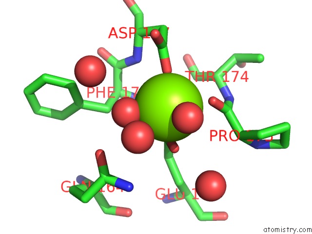
Mono view
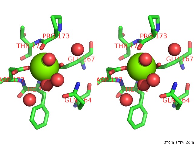
Stereo pair view
Mono view
Stereo pair view
A full contact list of Magnesium with other atoms in the Mg binding
site number 1 of Crystal Structure of A. Fulgidus Periplasmic Binding Protein Moda with Bound Tungstate within 5.0Å range:
|
Reference:
K.Hollenstein,
D.C.Frei,
K.P.Locher.
Structure of An Abc Transporter in Complex with Its Binding Protein. Nature V. 446 213 2007.
ISSN: ISSN 0028-0836
PubMed: 17322901
DOI: 10.1038/NATURE05626
Page generated: Wed Aug 14 01:32:05 2024
ISSN: ISSN 0028-0836
PubMed: 17322901
DOI: 10.1038/NATURE05626
Last articles
Zn in 9MJ5Zn in 9HNW
Zn in 9G0L
Zn in 9FNE
Zn in 9DZN
Zn in 9E0I
Zn in 9D32
Zn in 9DAK
Zn in 8ZXC
Zn in 8ZUF