Magnesium »
PDB 2ouq-2p88 »
2our »
Magnesium in PDB 2our: Crystal Structure of PDE10A2 Mutant D674A in Complex with Camp
Enzymatic activity of Crystal Structure of PDE10A2 Mutant D674A in Complex with Camp
All present enzymatic activity of Crystal Structure of PDE10A2 Mutant D674A in Complex with Camp:
3.1.4.17;
3.1.4.17;
Protein crystallography data
The structure of Crystal Structure of PDE10A2 Mutant D674A in Complex with Camp, PDB code: 2our
was solved by
H.C.Wang,
Y.D.Liu,
J.Hou,
M.Y.Zheng,
H.Robinson,
with X-Ray Crystallography technique. A brief refinement statistics is given in the table below:
| Resolution Low / High (Å) | 30.00 / 1.45 |
| Space group | P 21 21 21 |
| Cell size a, b, c (Å), α, β, γ (°) | 49.296, 82.285, 153.999, 90.00, 90.00, 90.00 |
| R / Rfree (%) | 21.8 / 23.6 |
Magnesium Binding Sites:
The binding sites of Magnesium atom in the Crystal Structure of PDE10A2 Mutant D674A in Complex with Camp
(pdb code 2our). This binding sites where shown within
5.0 Angstroms radius around Magnesium atom.
In total 2 binding sites of Magnesium where determined in the Crystal Structure of PDE10A2 Mutant D674A in Complex with Camp, PDB code: 2our:
Jump to Magnesium binding site number: 1; 2;
In total 2 binding sites of Magnesium where determined in the Crystal Structure of PDE10A2 Mutant D674A in Complex with Camp, PDB code: 2our:
Jump to Magnesium binding site number: 1; 2;
Magnesium binding site 1 out of 2 in 2our
Go back to
Magnesium binding site 1 out
of 2 in the Crystal Structure of PDE10A2 Mutant D674A in Complex with Camp
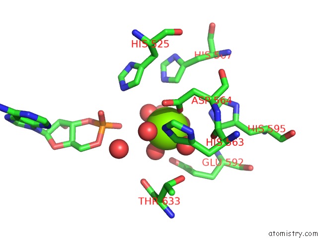
Mono view
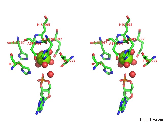
Stereo pair view
Mono view
Stereo pair view
A full contact list of Magnesium with other atoms in the Mg binding
site number 1 of Crystal Structure of PDE10A2 Mutant D674A in Complex with Camp within 5.0Å range:
|
Magnesium binding site 2 out of 2 in 2our
Go back to
Magnesium binding site 2 out
of 2 in the Crystal Structure of PDE10A2 Mutant D674A in Complex with Camp
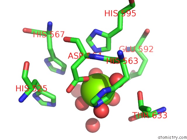
Mono view
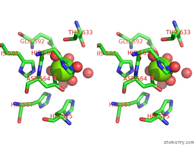
Stereo pair view
Mono view
Stereo pair view
A full contact list of Magnesium with other atoms in the Mg binding
site number 2 of Crystal Structure of PDE10A2 Mutant D674A in Complex with Camp within 5.0Å range:
|
Reference:
H.Wang,
Y.Liu,
J.Hou,
M.Zheng,
H.Robinson,
H.Ke.
From the Cover: Structural Insight Into Substrate Specificity of Phosphodiesterase 10. Proc.Natl.Acad.Sci.Usa V. 104 5782 2007.
ISSN: ISSN 0027-8424
PubMed: 17389385
DOI: 10.1073/PNAS.0700279104
Page generated: Wed Aug 14 01:57:27 2024
ISSN: ISSN 0027-8424
PubMed: 17389385
DOI: 10.1073/PNAS.0700279104
Last articles
Zn in 9JYWZn in 9IR4
Zn in 9IR3
Zn in 9GMX
Zn in 9GMW
Zn in 9JEJ
Zn in 9ERF
Zn in 9ERE
Zn in 9EGV
Zn in 9EGW